DETERMINATION OF AMINOGLYCOSIDES IN FOOD BY FLUORESCENCE POLARIZATION IMMUNOASSAY

Фарафонова О.В.1, Васильев С.В.2, Еремин С.А.3, Ермолаева Т.Н.4
1Кандидат химических наук, 2Студент, 4Доктор химических наук, Липецкий государственный технический университет; 3Доктор химических наук, Московский государственный университет имени М.В. Ломоносова
ОПРЕДЕЛЕНИЕ АМИНОГЛИКОЗИДНЫХ АНТИБИОТИКОВ В ПИЩЕВЫХ ПРОДУКТАХ МЕТОДОМ ПОЛЯРИЗАЦИОННОГО ФЛУОРЕСЦЕНТНОГО ИММУНОАНАЛИЗА
Аннотация
Разработаны методики определения аминогликозидных антибиотиков (гентамицин, канамицин, стрептомицин, амикацин, неомицин) в пищевых продуктах методом поляризационного флуоресцентного иммуноанализа (FPIA). Исследовано влияние размера и структуры флуоресцентной молекулы на степень поляризации флуоресценции. Рассчитаны константы трейсеров и аффинности поликлональных антител, оптимизированы рабочие концентрации трейсеров и антител, обеспечивающие максимальное значение аналитического сигнала. Методики апробированы для анализа куриного мяса, яиц, молока.
Ключевые слова: поляризационный флуоресцентный иммуноанализ, антибиотик, гентамицин, канамицин, амикацин, неомицин, стрептомицин, пищевой анализ.
O.V. Farafonova1, S.V. Vasiliev 2, S.A. Eremin3, T.N. Ermolaeva4
1Candidate of Chemistry Sciences, 2Student, 4Doctor of Chemistry Sciences, Lipetsk State Technical University; 3S.A. Eremin, Lomonosov Moscow State University
DETERMINATION OF AMINOGLYCOSIDES IN FOOD BY FLUORESCENCE POLARIZATION IMMUNOASSAY
Abstract
The methodic for quantitative determination of aminoglycoside antibiotics (gentamicin, kanamycin, streptomycin, amikacin, neomycin) in food by polarization fluorescent immunoassay (FPIA) is developed. The size and structure influence of a fluorescent molecule on a fluorescence polarization degree is analyzed. Affinity constants of antibodies to compounds and tracers were estimated, optimized working concentration of tracers and antibodies that provide the maximum value of analytical signal. Methods were tested in the antibiotics identification in milk, eggs and chicken.
Keywords: polarization fluorescence immunoassay, antibiotic, gentamicin, kanamycin, amikacin, neomycin, streptomycin, food testing.
INTRODUCTION
The regular use of aminoglycoside antibiotics for the treatment and prevention of cattle, poultry and bees infectious diseases leads to the accumulation of these substances in the muscle tissues of animals, milk, honey, eggs. This greatly complicates the preventive measures and treatment of diseases due to the toxic effect on cells and tissues, allergic reactions occurrences, antibiotic-resistant microorganisms appearance.
There are many methods for aminoglycoside antibiotics determination because of their widespread use. Such as: microbiological and physico-chemical methods. Chromatographic methods require long sample preparation, which includes separation and partition of components [1 - 4]. Microbiological methods have low specificity and selectivity [5 – 7]. Fluorescence Polarization Immunoassay (FPIA) can avoid these shortcomings. FPIA is a homogeneous method based on competition between defined antigen and the fluorescent-labeled antigen, for a limited number of specific antibodies binding sites. FPIA uses fluorescence polarization as the measurable value, which depends on the mobility of a fluorescent molecule. This allows you to control the binding of low-molecular labeled antigen, called tracer, with high-molecular antibodies. The measurement is performed on standard equipment, using mono and polyclonal antibodies. Polyclonal antibodies are used more frequently [8].
There are FPIA determination methods of gentamicin, kanamycin, amikacin, streptomycin in medical objects: plasma and serum [9 - 14]. These methods are means of regulating the dosage and duration of treatment, which help therapists. Plasma and serum may contain interfering components: proteins, lipids, bilirubin and hemoglobin [15, 16]. At the same time you can not apply these techniques to determine the food, because it is necessary to analyze the influence of the food matrix. Determination of aminoglycosides in food is a topical issue in society.
Maximum permissible concentration of aminoglycosides is different and depends on the type of antibiotic and determined object, for many aminoglycosides this concentration has not even entered. MPC of streptomycin is 500 µg/kg in meat and liver, 1000 µg/kg in kidney, 200 µg/l in milk. In Germany, MPC of streptomycin in honey is 20 µg/kg. MPC of gentamicin and kanamycin in various food products is 100 µg/kg, neomycin 500 µg/kg.
MATERIALS AND METHODS
Chemicals. Azide, sodium chloride, acetonitrile, methanol, ethanol, chloroform, acetone (high purity grade, "Himmed", Russia), triethylamine (chemically pure grade “Merck”, Germany), dimethylformamide (brand purity, "Vekton”, Russia). Streptomycin, gentamicin, kanamycin, amikacin, neomycin; digidrostreptomicyn bought in Abkame and used according to manufacturer's instructions. Polyclonal antibodies to streptomycin (An-STP), gentamicin (An-GENT), kanamycin (An-KANA), kindly provided by Professor R. Abukneshey (Imperial College, UK). Neomycin, gentamicin (Abcam) fluorescein isothiocyanate (FITC), cyanuric chloride (CC), N-hydroxysuccinimide (NHS), N, N'-dicyclohexylcarbodiimide (N, N'-DCC) (chemically pure grade, «Sigma Aldrich Corporation », USA), ethylenediamine dihydrochloride (grade chemically pure," Himmed ", Russia); tracers - to gentamicin with a fluorescent label fluorescein isothiocyanate (GENT - FITC), 5 - ([4,6-dihlorotriazin-2-yl] amino)-fluorescein (GENT - DTAF), to streptomycin with fluorescent label Fluoresceinthiocarbamyl ethylenediamine (STR-cc - EDF) ("Sigma", USA).
To prepare 1 liter of borate buffer solution, pH = 8.0 was dissolved 0,95g of Nа2В4О7 10Н2О and 1,00g Na3N.
To select antibiotics labeled with a fluorescent label, and their purification by thin layer chromatography were used «Silufol» (Czech Republic) plates.
Synthesis of the tracers
Method 1. The synthesis of tracers for streptomycin, dihydrostreptomycin, kanamycin, amikacin, gentamicin was carried out by the following procedure: 15 mg of antibiotic was dissolved in 500 µg of water-methanol mixture (3:1) containing 30 µL of triethylamine. 2.5 mg FITC was dissolved in 300 µL of methanol containing 10 mL of triethylamine. After that the first solution with mixing was added dropwise to a second solution. The reaction mixture was stirred for 12 hours in darkness at ambient temperature.
Cleaning tracers by thin layer chromatography. The reaction mixture containing the tracer, the initial components, byproducts of the reaction and decomposition of the tracer, as well as auxiliary reagents applied to the "Silufol" (Czech Republic) plate, size 2020 cm: the developer - a mixture of methanol: chloroform 1:4 (by volume). The stripes, painted in yellow, and showed up in ultraviolet light contained fluorescent compounds with different values of Rf, were collected from the plate and eluted in 1 ml of methanol. The purification of fluorescent compounds monitored chromatographically by the values of Rf = 0.1, and further in work we used only those tracers, which bound to a specific antibody. Methanol solution of tracer was stored at 4oС, in work tracers solutions further diluted with borate buffer solution to a working concentration.
Method 2. In the synthesis of NEOM-FITC tracer used 80 mg of antibiotic. The rest of the technique is similar to the Method 1.
The fluorescence polarization was measured at TDx-analyzer ("Abbott", USA) and the polarization fluorimeter «Beacon 2000» («Panvera», USA).
For antibody titration curves were carried out serial dilution of tested antibodies solutions: in 10 cuvettes successively diluted antibody of 500 µL borate buffer solution, starting at a dilution of 1:20. Then in all cuvettes were added 500 µL of tracer working solution and measured the fluorescence polarization (mP). According to the results of measurements were build “antibody dilution” – “value of fluorescence polarization” (mP) graph, by this graph were determined the dilution of antibody that causes 70% binding of tracer.
Measurements on the TDx device in semi-automatic mode with manual dosing were performed by the “Photo Check” program. Within calibration curve building, to 50 µL of antibiotic solution with a concentration of 0.001 - 100.0 µg / mL were added 500 µL of tracer working solution and 500 µL antibodies with the dilution corresponding to 70% binding. The mixture was stirred and measured the fluorescence polarization. Denatured proteins were separated on a desktop centrifuge.
Sample preparation
Preparation of samples of chicken meat carried by the following method: the meat pre-homogenized, then extracted with an antibiotic. To 10,0 g sample was added 50 ml water and 10 ml of saturated solution of (NH4)2SO4, for the separation of protein components, stirred, cooled and centrifuged for 8 min (7000 rpm). The supernatant was used for analysis.
Milk samples before analysis were centrifuged for 3 minutes (6000 rpm), then removes the top layer of fat and the remaining skim milk was subjected to one of two types of processing. In the first case to 500 ml of milk was added 500 ml (or 1 ml) of methanol, ethanol, acetonitrile to precipitate the milk proteins. In the second case, add a saturated solution of ammonium sulfate. Then process the samples were centrifuged for 5 minutes (9000 rpm), supernatants were collected and conducted its analysis.
Chicken eggs were preparing for the procedure: Samples were homogenized eggs, were collected 2.0 g of the obtained mixture, to which was added 10 Pl antibiotics (1 mg/ml) and 2.0 ml of acetonitrile, ethanol, methanol, stirred and centrifuged for 2 min (8000 rpm min). The supernatant portion was separated and evaporated; the residue was dissolved in 980 ml buffer solution.
RESULTS AND DISCUSSION
The first phase of the designed FPIA methodology is getting its fluorescent derivative, called a tracer. Aminoglycoside antibiotics contain active amino groups, which can easily react with FITC fluorescent label. Have been synthesized six tracers for different antibiotics (gentamicin, kanamycin and streptomycin, digidrostreptamycin, amikacin, neomycin) with the same fluorescent label GENT-FITC, KANA-FITC, STP-FITC, DSTP-FITC, AMIC-FITC and NEOM-FITC, respectively. Antibiotic molecules are very similar to each other. The difference is only in the number of substituents in the ring, as well as in the structure of neomycin. For all antibiotics except neomycin, used the first method of synthesis. The synthesis of neomycin, since a large number of active functional groups, used the second method, where excess of antibiotic exclude the binding of all NH2-groups. Also, the study takes into account the tracers possible cross-reaction and experiments conducted with various specific antisera to antibiotics.
The sensitivity and efficiency FPIA depends on the correct choice of the pair immunoreagents: tracer - antibodies. Therefore, all received tracers were tested for binding to the corresponding specific antisera. To study the binding of antibody to antibiotic - fluorescent tracer were obtained antibodies dilution curves. These curves show a signal dependency from the dilution of antibody at constant concentration of tracer (Fig. 1).
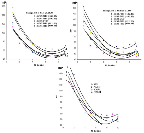
Fig. 1 - Dilution curves for antibodies with tracers
An important condition for the polarization of fluorescence immunoassay is a strong possibility of the immune complexes formation between the analyte and the antibody (KAf) and the tracer and antibody (KT). It is essential that KAf> KT more than 1-2 orders, because only in this case perhaps tracer ousting from a pre-organized complex. It was found that the binding constants of tracer-antibody and antibody-antigen of all applicable immunoreagents pairs differ on one order (Table 1), so all the studied immunoreagent could be used in FPIA.
High values of КАf about 108 showed high specificity and activity immunoreagents binding.
Table 1 - The affinity constants of antibody and tracer of applied immunoreagents pairs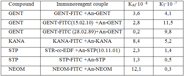
To assess cross-reactivity of polyclonal antibodies obtained to antibiotics were applied, evaluated the cross-response coefficient (IC, %) (Table 2).
Table 2 - Ratios of cross-reaction (IC, %)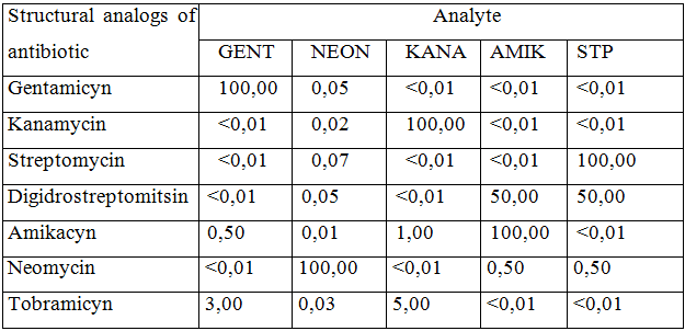
The values of these coefficients for the structural analogues are 0,01 - 5,00%, which indicate the possibility of high specific determination of individual compounds in the joint presence. An exception is the antibody to streptomycin, which exhibit a high cross-reactivity to its dihydro-derivative (50%).
Determined the reproducibility of the method both during one experiment and between several days. The coefficient of variation has not exceeded 3% in both cases.
The correctness of determination of antibiotics was tested by the “added – found” method (Table 3). Statistical analysis of the results using the Student criterion revealed no systematic errors. Low values of sr indicate high reproducibility of analysis.
Table 3 - Verifying the correctness of streptomycin determination (n = 3, P = 0,95)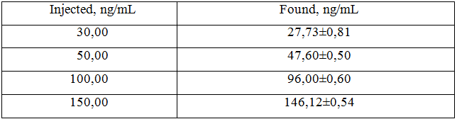
The developed methods of antibiotics definition applied to the analysis of modelling solutions. Metrological characteristics of definition are showed in table 4.
Table 4 - Metrological characteristics of antibiotics determination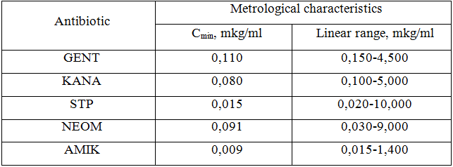
This data shows that the least limit of detection (9 ng/ml) characterizes FPIA method for amikacin. The study of specificity showed that the method FPIA using these antibodies have high enough specificity to identify each of the antibiotics in the presence of others.
Practical application FPIA when analyzing food.
Determination in milk. Determination in milk provided with specific samples training, based on the deposition of milk proteins. As the precipitants reagents were tested: methanol, ethanol, acetonitrile and a saturated solution of ammonium sulfate (NH4)2SO4. After analysis of samples compared precipitators was performed on the basis of fluorescence polarization values obtained for each antibiotic.
To streptomycin and amikacin was best precipitator methanol CH3OH. To this reagent in both cases the optimal ratio is between the values of mP 0 ng·ml-1 and a relative decrease of the analytical signal.
For streptomycin the technique has appeared enough sensitive as the final limit of detection of this preparation has appeared below its maximum concentration limit of 200 ng·ml-1. To evaluate the applicability of the developed techniques FPIA for analysis of real milk samples were carried out tests on the discovery of these drugs by the method “added – found” method.
Used milk is not supported none of the identified compounds, than in the samples of this milk were introduced known concentrations of a given antibiotic. Then the samples containing antibiotics were subjected to sample preparation with ammonium sulfate, and samples with streptomycin and amikacin were treated with methanol as reagent-precipitator (Table 5).
As can be seen from the table, extraction percentages for all antibiotics were more than 80%. Incomplete discovery of drugs in milk, probably due to the losses of the test compound in the process of sample preparation of milk: during extraction. The values obtained for each antibiotic showed the effectiveness of the developed techniques FPIA and their suitability for the analysis of real milk samples.
Table 5 - The measurement of antibiotics in milk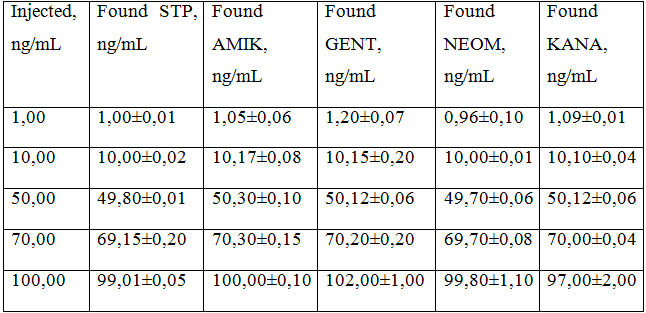
Determination in eggs. Chicken eggs were preparing applied the nonsolvent methanol, as with others nonsolvents, such as: acetonitrile, ethanol, methanol testimony polarization were higher than 150.
Determination of antibiotics in chicken meat. Explored ways of extracting antibiotics from chicken meat with acetonitrile [12 – 13], a mixture of acetonitrile with hexane and dichloroethane, aqueous solution of acetone, as well as ammonium sulfate solution (18%). It is shown that only when extracting an aqueous solution of antibiotics (NH4)2SO4 achieved almost complete removal of drugs (93% and above). In other cases, the extraction percentage is 60 - 80%.
The developed methods were tested in the chicken meat analysis. The content of antibiotic in the samples recalculated to 1 kg of product. In all cases, not exceeded the MCL values for streptomycin, but found too high content of gentamycin compared with the MCL (0.10 mg / kg), introduced for the European Union (in Russia the same indicator is absent).
Table 7 - Determination of antibiotics in food (P = 0,95, n = 3)
Studies have shown that the results of the determination of antibiotics in food products are characterized by high reproducibility, selectivity and accuracy, so the technique can be recommended for monitoring food safety and trace concentrations detections of aminoglycoside antibiotics in meat and milk.
References
- Bruijnsvoort M., Ottink S.J.M., Jonker K.M, Boer E. Determination of streptomycin and dihydrostreptomycin in milk and honey by liquid chromatography with tandem mass spectrometry // M. Bruijnsvoort, S.J.M. Ottink, K.M. Jonker, E.Boer // J.Chromatogr.–2004.–V. 1–2.–P. 137–142.
- Lima J., Delerue – Martos C., Vaz C. Enzymatic determination of choline in milk using a FIA system with potentiometric detection // Analyst.– 2000.– V. 125.– P. 1281 – 1284.
- Kaufmamn A., Maden K. Determination of 11 Aminoglycosides in Meat and liver by liquid chromatography with tandem mass spectrometry // Journal of AOAC International. –2005.– V. 88. –P. 1118–1125.
- Abuknesha Ramadan A., Luk Connie Enzyme immunoassays for the analysis of streptomycin in milk, serum and water: development and assessment of a polyclonal antiserum and assay procedures using novel streptomycin derivatives // Analyst – 2005. - V.130.-No 6. - P. 964-970.
- Haasnoot W., Stouten P., Cazemier G., Lommen A., Nouws J. F. M., Keukens H. J. Immunochemical detection of aminoglycosides in milk and kidney // State Institute for Quality Control of Agricultural Products (RIKILT-DLO), Bornsesteeg 45.-1999.
- Jin Y., Jang J. K., Han C. H., Lee M. H. J. Development of ELISA and immunochromatographic assay fort he detection on gentamicin // Vet Sci. – 2006. - No 7. – P. 111-117.
- Dang P. K., Degand G., Douny C., Ton V. D., Maghuin-Rogister G., Scippo M-L. Optimisation of a new two-plate screening method for the detection of antibiotic residues in meat // International Journal of Food Science and Technology – 2011. – V. 46. – P. 2070 .
- Zhang S., Wang Z., Nesterenko I.S., Eremin S. A., Shen J. Fluorescence Polarization Immunoassay based on a Monoclonal Antibody for the Detection of Sulfamethazine in chicken muscle // J. Food Sci. Tech. – 2007. –V.42.– P. 36-44.
- Tsuruoka M., Karube I. Rapid Hybridization at High Salt Concentration and Detection of Bacterial DNA Using Fluorescence Polarization // Chem. High T. SCR. – 2003. – V. 3. – P. 225-234.
- Goryacheva I.Yu., Saeger S.De, Nesterenko I.S., Eremin S.A., Peteghem C. Van. Rapid all-in-one three-step immunoassay for non-instrumental detection of ochratoxin A in high-coloured herbs and spices // Talanta. – 2007. –V.72. – P.1230-1234.
- Wang Z., Zhang S., Nesterenko I.S., Eremin S.A., Shen J. Fluorescence Polarization Immunoassay for Sulfamethoxypyridazine and Sulfachloropyridazine // Agric. Food Chem. – 2007. – V. 55. – P. 6871-6878.
- Eremin S.A., Smith D.S. Fluorescence Polarization Immunoassays for Pesticides // Comb. Chem. High T. Scr. - 2003. - V. 6. - P. 79-99.
- Eremin S.A., Murtazina N.R., Ermolenko D.N., Zherdev A.A., Mart'ianov A.A., Yazynina E.V., Michura I.V., Formanovsky A.A., Dzantiev B.B. Production of polyclonal antibodies and development of fluorescence polarization immunoassay for sulfanilamide // Anal. letters.- 2005. - V. 38. - P. 951-969.
- Smith D. S., Eremin S. A. Fluorescence polarization immunoassays and related methods for simple, high-throughput screening of small molecules // Bioanal. Chem. - 2008. - V. 391.- P. 1499-1507.
- Maragos C.M., Kim E.-K. Detection of zearalenone and related metabolites by fluorescence polarization immunoassay // Food Protec.-2004.- V.6 (5).- P. 1039-1043.
- Do E.U., Choi G., Shin J., Jung W.-S., Kim S.-I Fluorescence polarization assays for high-throughput screening of neuropeptide FF receptors // Analytical Biochemistry. – 2004. - V. 33. – No 1. – P. 156-163.