THE EXPERIMENTAL MODEL OF ACUTE PANCREATITIS

Рахимов Р.Р.
ORCID: 0000-0002-2488-597X, Аспирант, ФГБОУ ВО Башкирский государственный медицинский университет МЗ РФ
ЭКСПЕРИМЕНТАЛЬНАЯ МОДЕЛЬ ОСТРОГО ПАНКРЕАТИТА
Аннотация
В данной работе представлена экспериментальная модель острого панкреатита с формированием внутреннего панкреатического свища. Воспроизведён некроз перешейка поджелудочной железы у 5 поросят «мясной» породы. Способ основан на травматизации поджелудочной железы сосудистым раздавливающим зажимом типа Бильрот, введением 1 мл аутожелчи и потреблением 40 % раствора этилового спирта для стимуляции секреции поджелудочной железы. Развитие острого панкреатита было подтверждено лабораторным, иммуногистохимическим и патогистологическим исследованиями. Достигнута отечно-геморрагическая стадия острого панкреатита, сменившееся геморрагическим панкреонекрозом. Данная экспериментальная модель может использоваться в научно-исследовательских лабораториях для изучения этиопатогенеза формирования внутреннего панкреатического свища и выработки тактики лечения острого панкреатита.
Ключевые слова: экспериментальная модель, острый панкреатит, внутренний панкреатический свищ.
Rakhimov R.R.
ORCID: 0000-0002-2488-597X, Postgraduate student, Bashkir State Medical University
THE EXPERIMENTAL MODEL OF ACUTE PANCREATITIS
Abstract
This paper presents an experimental model of acute pancreatitis with the formation of an internal pancreatic fistula. Isthmus reproduced necrosis of the pancreas in 5 pigs meat of the breed. The method is based on trauma of the pancreas by vascular clamp type Billroth, introducing 1 ml auto bile and consumption 40% ethyl alcohol solution for stimulation of pancreatic secretion. The development of acute pancreatitis was confirmed by laboratory, immunohistochemical and histopathological examination. Reached edematous hemorrhagic stage of acute pancreatitis changed to hemorrhagic necrotizing pancreatitis. This experimental model can be used in research laboratories for the study of the pathogenesis of the disconnected pancreatic duct syndrome and policy-making treatment of acute pancreatitis.
Keywords: experimental model, acute pancreatitis, disconnected pancreatic duct syndrome.
Introduction
Acute pancreatitis is one of the most serious diseases in emergency abdominal surgery. Despite progress in improving diagnosis, intensive antibiotic therapy and surgical treatment options involving minimally invasive surgery, the overall mortality in severe acute pancreatitis in recent decades is maintained at a high level up to 10-30% and reaches at 85% infected pancreatic necrosis [1,P.444],[2, P. 245]. Today etiological aspects of acute pancreatitis are not well understood.
Currently, the different groups of playback methods of acute pancreatitis: toxic-infectious, traumatic, vascular, allergic, canalicular-hypertensive model.
Many models of acute pancreatitis have lost their relevance. There is need for a new experimental model of acute pancreatitis, which is closest to the clinical picture. Not one of the proposed methods do not reflect the impact of destructive inflammatory processes taking place in the pancreas towards peripancreatic and retroperitoneal cellular spaces [3, P.443].
Dyuzheva TG et al. described the internal pancreatic fistula during deep pancreatic necrosis isthmus to damage the main pancreatic duct, pancreatic juice in which the enzymes produced by the body and the tail reaches the duodenum. Lymphatic vessels carry draining function is observed as a result of impregnation of the retroperitoneal fat pancreatic juice [4, P.94]. This condition is accompanied by severe endotoxemia development of inflammatory processes in peripancreatic and retroperitoneal cellular spaces.
Aim of the study is to establish an experimental model of acute pancreatitis with necrosis of the isthmus of the pancreas followed by the formation of internal pancreatic fistula.
Materials and methods
The experiment was performed at the Department of Morphology, pathology, pharmacy and non-communicable diseases Bashkir State Agrarian University in cooperation with the staff of the departments of pathological anatomy and of surgical diseases with the course of colorectal surgery BSMU. The studies were conducted under the protection of animals at the convention (№125 from 13.11.1987) and were approved by the ethical committee of Bashkir State Medical University.
The experimental study was performed on 10 pigs' meat "of the breed, both sexes, average body weight 10 ± 0,5 kg. When selecting animal laboratory for experimental research, we proceeded from the assumption that pigs are very similar to the man on the anatomical and physiological characteristics of the digestive tract and abdominal organs. Pigs were divided into two groups of 5 animals. The first group - the control. Animals with the unmodified state of the pancreas and retroperitoneal fat were included. The second group - experimental. Care for the animals carried out under the same conditions with three meals a day and constant access to water. Stop feeding pigs 12 hours before the start of the pilot studies, and water for 6 hours. Perform blood sampling from the marginal ear vein for morphological studies prior to, during and at the end of the experiment. Premedication performed 15 minutes before induction of anesthesia. We used a combination of atropine and 0.1% dimedrol 0.1 mg / kg intramuscularly. Anesthesia was carried out drug benefit "Zoletil 100" 15 mcg / kg intramuscularly. Miorelaxation performed xylazine 4.5 mg / kg. Animal awakens from sleep medication caffeine sodium benzoate 20% -1 ml.
To create an internal pancreatic fistula model was developed with traumatic pancreatic isthmus and the introduction autobile in damage zone. Made Upper midline laparotomy for abdominal white line by the standard technique. Imposes a purse string suture in the bottom of the gallbladder, where it carried out a puncture with bile fence. Shred bag stuffing. It exposes the pancreas. Vascular crushing clamp type Billroth performed trauma in the area of the isthmus of the pancreas. Injected 1 ml autobile in trauma area in figure1. Sutured wound of the anterior abdominal wall in layers.
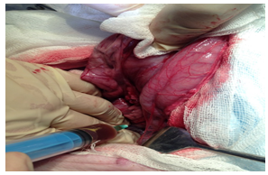
Fig. 1 - Introduction of bile in the isthmus of the pancreas
In order to stimulate the secretion of pancreatic animals drinking water three times from 40% ethanol solution. After drinking pigs were flaccid, adynamic, refused to feed. By the end of the third day after the experimental model of acute pancreatitis producing animals withdrawn from the study. The first step was introduced into the anesthetic drug zoletil 100, then infused intravenously 10% solution of lidocaine in the marginal ear vein. In carrying out investigations to observe the "rules of work with the use of experimental animals," according to the order of Ministry of Health of the USSR № 755 from 12.08.77
The results of experimental studies
All 10 animals were conducted laboratory tests and ultrasound SSB veterinary ultrasound scanner ECOSON 700W. In the main group in the KLA a marked leukocytosis 16,7 ± 2,3 h109 / l with a shift to the left leukocyte, ESR 45,2 ± 4,2 mm / h. However, in the control group to 14.2 ± KLA leukocytes 2,3h109 / l. In the main group in the biochemical analysis of blood amylase stood at 2634,5 ± 134,5 u / l. The maximum value of blood amylase was 2894 units. / L. In the control group, the blood amylase was 247.5 ± 3.1 u / l. The maximum value was 253 u / l. 109 / L.
Spend anatomical and pathological analysis of the changes in the internal organs in the simulation of internal pancreatic fistula with acute severe pancreatitis. Local changes of the pancreas and retroperitoneal fat were evaluated by immunohistochemistry and histopathological examination. almost no free liquid was in the abdominal cavity. However, in the packing bag was found hemorrhagic effusion. A marked swelling of the pancreas with pockets of fat necrosis on the surface. The Isthmus of pancreatic necrosis zone area is defined with hemorrhagic impregnation of pancreatic tissue damage the main pancreatic duct in figures 2 and 3.
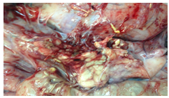
Fig.2 - There are pancreatic edema and parapancreatic tissue with foci of fat necrosis and hemorrhagic impregnation
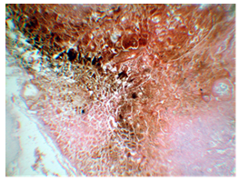
Fig. 3 - Hemorrhagic necrosis zone and impregnating the isthmus in the pancreas. H & E stain, SW.100
There was migration of leukocytes from pancreatic tissue into the lumen of the main pancreatic duct, pronounced swelling of the duct obstructed outflow of pancreatic juice, which indicated the presence of internal pancreatic fistula in figure 4.
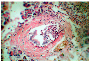
Fig. 4 - Migration of leukocytes from cancer tissue into the lumen of the main pancreatic duct at the isthmus of pancreatic necrosis. H & E stain, SW.400
The left half of the retroperitoneal fat, together with the left perirenal fat were swollen. With the opening of the data space is allocated in a transparent fluid, also expressed vascular congestion was found. The content of amylase in the exudate was 1025,3 ± 125,3 u / l in the main group. However, in the control group retroperitoneal fat was unchanged.
Discussion
Currently, there are a large number of experimental models of acute pancreatitis. Each model reflects different chain of pathogenetic processes caused by different triggers. These experimental models are technically difficult to implement. For example, the required ligation of the main pancreatic duct or the need for accurate dosing damaging drug. Vascular-allergic models are very difficult to achieve and reflect the real picture of the disease. Not one of the previously proposed models do not reproduce isolated necrosis of the isthmus of the pancreas [5, P.205], [6, P.186].
As a result, the new model has been developed acute pancreatitis. These laboratory tests, macro and microscopic lesions in the pancreas and retroperitoneal spaces confirm the development of acute pancreatitis with an internal pancreatic fistula in animals. This model reproduces the clinical picture of acute pancreatitis in patients.
Conclusions
The proposed method is an experimental model of acute pancreatitis allows most closely reflect the pathogenesis of pancreatic fistula formation of the internal in humans with acute pancreatitis and changes in the retroperitoneal fat. This model is available to run and can be used in research laboratories for the study of the etiology, pathogenesis of the disease and finding his new treatments.
Список литературы / References
- Effects of Castanospermine on Inflammatory Response in a Rat Model of Experimental Severe Acute Pancreatitis./Hong YP, Chen C, Guo WY, Zhao L, Mei FC and others //Arch Med Res.- 2016 - №6. - P.436-445. doi: 10.1016/j.arcmed.2016.11.007.
- Clusterin and Pycr1 alterations associate with strain and model differences in susceptibility to experimental pancreatitis./ Iyer S, Park MJ, Moons D, Kwan R and others //Biochem Biophys Res Commun. - 2016 - Vol.16 - №6. doi: 10.1016/j.bbrc.2016.12.039.
- Effects of Castanospermine on Inflammatory Response in a Rat Model of Experimental Severe Acute Pancreatitis. /Hong YP, Chen C, Guo WY, Zhao L, Mei FC and others // Arch Med Res. – 2016. - №6. – P.436-445. doi: 10.1016/j.arcmed.2016.11.007.
- Configuration pancreatic necrosis and differentiated treatment of cancer and the differentiated treatment of acute pancreatitis /Dyuzheva TG, Juice E. Schaefer AV Akhaladze G.G. and others // Annals of Surgical hepatology. -2013. – Vol. 18. - №1. - 92-102.
- Reduced Pancreatic Exocrine Function and Organellar Disarray in a Canine Model of Acute Pancreatitis / Yuepeng Jin, Yongyu Bai, Qiang Li, Pravin Avinash Bhugul and others //PLoS One. – 2016. – Vol.11. - №2. URL:http://journals.plos.org/plosone/article?id=10.1371/journal.pone.0148458
- Effects of everolimus on a rat model of cerulein-induced experimental acute pancreatitis / Alper Bilal Özkardeş, Birkan Bozkurt, Ersin Gürkan Dumlu, Mehmet Tokaç and others //Ulus Cerrahi Derg. – 2015. – Vol.31. - №4. –P.185-191.