Вернуться к статье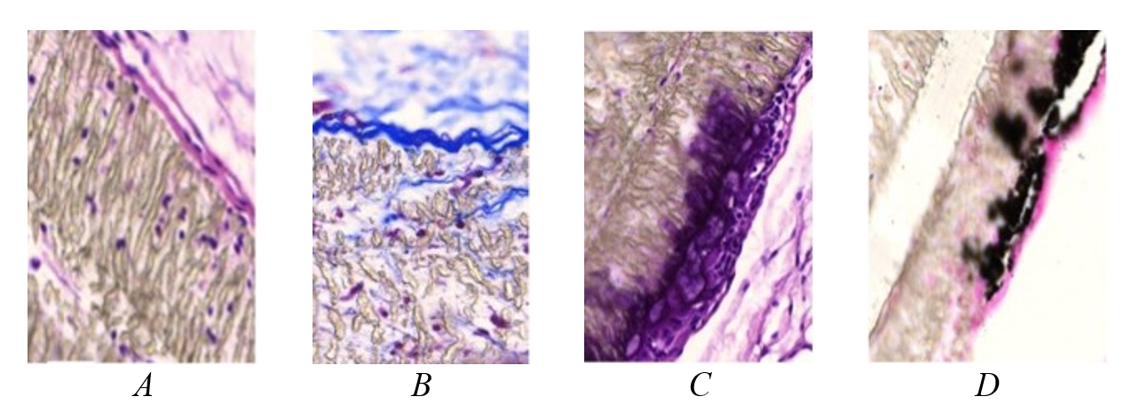
Морфология зоны имплантации синтетических герниопротезов, модифицированных углеродом
Figure 1 - 180 days after implantation of an uncoated polytetrafluoroethylene plate:
A – the implant structure is moderately loosened; on the outside, single loosened collagen fibers of the connective tissue of the anterior abdominal wall1; B – leukocyte migration into the pores of the material, ingrowth of single thin collagen fibers2; C – giant foreign body cell layer3; D – area of calcification in the region of foreign body cell accumulation4
1 – stained with hematoxylin and eosin; microphoto. ×100; 2 – mallory staining; microphoto. ×200; 3 – stained with hematoxylin and eosin; microphoto. ×100; 4 – kossa impregnation; microphoto. ×100