Современные диагностические подходы к выявлению гепатопатий у производственных животных
Современные диагностические подходы к выявлению гепатопатий у производственных животных
Аннотация
На фоне активного расширения производства молочной продукции и повышения продуктивности сельскохозяйственных животных, благодаря инновационным разработкам в селекционно-племенном направлении, высокопродуктивные животные сталкиваются с проблемой развития патологий печени различной природы, связанных с регулярным превышением физиологических возможностей вида и нарушением адаптации индивидуумов к высокой интенсивности катаболических и анаболических процессов. В статье представлен обзор последних исследований отечественных и зарубежных специалистов по вопросу поиска и разработки наиболее эффективных и воспроизводимых методов диагностики патологий гепатобилиарной системы у продуктивных животных. Был проведен анализ статистических данных об объемах производства молока, а также проведена оценка взаимосвязи продуктивности животных с распространенностью заболеваний печени у высокопродуктивных коров. В статье рассматривается эффективность современных диагностических подходов, основанных на анализе показателей биохимического профиля, индикаторов эндогенной интоксикации и специфических биомаркеров, а также оценивается неинвазивный метод исследования печени — транзиторная эластография — с точки зрения интеграции в условия животноводческого комплекса. Следует отметить, что некоторые подходы требуют дополнительных исследований для установления их эффективности и воспроизводимости в клинических условиях.
1. Введение
Dairy farming is one of the most popular areas in the agricultural sector of modern Russia. The dynamic increase in the volume of dairy production by enterprises of the agro-industrial complex is due to the annual growth of consumer demand. The trend towards increasing cow's milk productivity is currently based on the principles of intensification and modernization of production processes, including improving the quality of feed resources, improving the technology of growing and keeping productive animals, introducing modern equipment, and deepening selection and breeding work .
According to the Unified Interdepartmental Information and Statistical System (UIISS), the average annual milk yield per cow in almost all categories of producers demonstrates positive dynamics (Fig. 1, Tab. 1). The consistent and planned development of domestic dairy cattle breeding and the sustainable increase in production volumes are also evidenced by statistical reports from specialists of the Federal State Budgetary Institution “Center for Agroanalytics” Yu. Tsindrina and V. Kartashova (Fig. 3) .
Table 1 - Dynamics of average milk yield per cow in farms of different categories
Year | Farms of all categories | Category of farm Peasant (farm) farms and individual entrepreneurs | Agricultural enterprises | Households |
2015 | 4 134 | 3 465 | 5 140 | 3 500 |
2016 | 4 218 | 3 499 | 5 370 | 3 484 |
2017 | 4 368 | 3 628 | 5 660 | 3 518 |
2018 | 4 492 | 3 689 | 5 945 | 3 463 |
2019 | 4 640 | 3 791 | 6 286 | 3 471 |
2020 | 4 839 | 3 979 | 6 728 | 3 471 |
2021 | 4 988 | 3 963 | 7 007 | 3 538 |
2022 | 5 194 | 3 989 | 7 440 | 3 572 |
2023 | 5 322 | 3 449 | 7 911 | 3 600 |
2024 | 5 518 | 3 482 | 8 290 | 3 630 |
Total production volumes for 2024, million tons | 16.8 | 1.4 | 10.4 | 5.0 |
100% | 8.2% | 62.1% | 29.7% |
Note: based on the source [2]
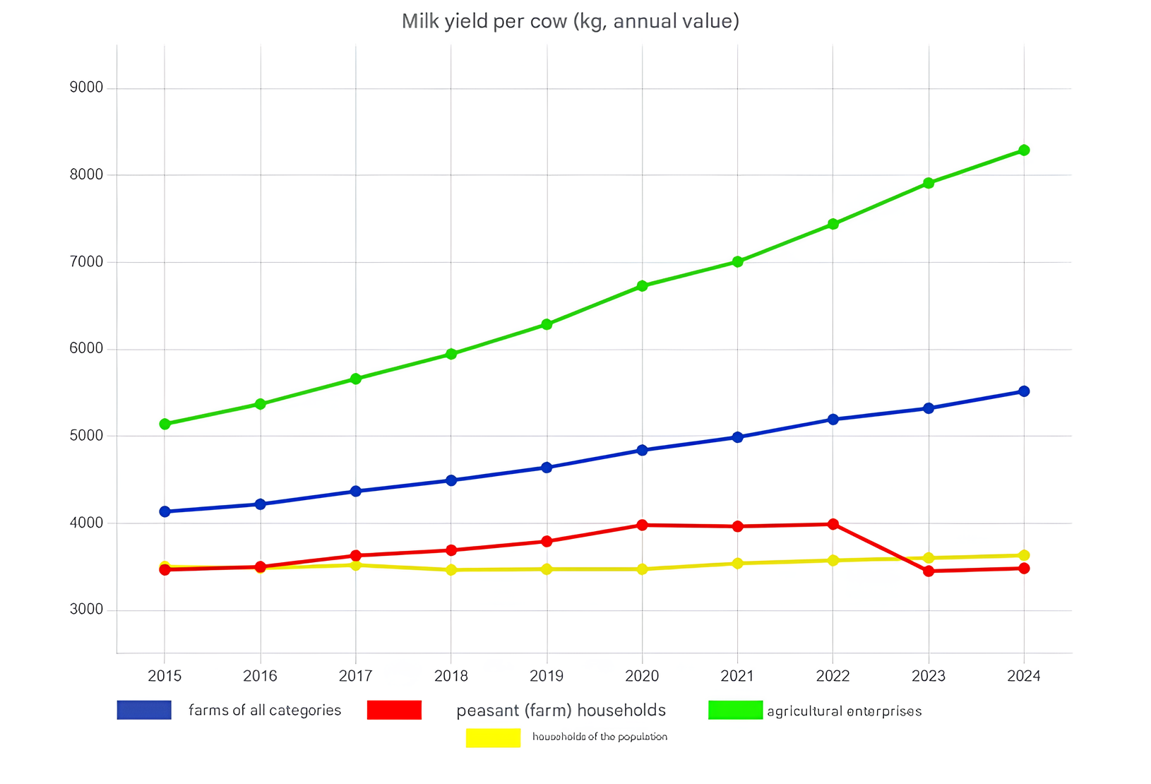
Figure 1 - Dynamics of average milk yield per cow in farms of different categories
Note: based on the source [2]

Figure 2 - Volumes of milk production in Russia according to the Federal State Budgetary Institution “Center for Agroanalytics”
Note: based on the source [3]
In a scientific work by Russian researchers, it was noted that approximately 45–80% of carcasses of forced-killed high-yielding Holstein-Friesian and Holsteinized Black-and-White cows, which annually produced more than 7 thousand liters of milk, had pathomorphological changes in the hepatobiliary system . Average productivity of this level is observed, for the most part, in large agricultural enterprises (Table 1). The economic damage from hepatopathies consists of a decrease in milk productivity of cows (on average, by 15–26%), a decrease in the rate of live weight gain (on average, by 10–15%), and the culling of every 8th- 10th liver when animals are slaughtered . The most common hepatopathies of high-yielding cows include hepatitis and fatty and granular dystrophy of the organ.
According to the results of studies conducted on the basis of livestock enterprises in the Belgorod, Lipetsk, Oryol, Voronezh, Moscow, Tver, and Tula regions, 47.8% of animals have liver diseases. A positive correlation was also found between the incidence rate and the productivity of individuals: 570 cows with a milk yield of 7000-8500 kg per lactation had a morbidity rate of 52.4%, and among 725 cows with a milk yield of 4500–5200 kg per lactation, this figure was 20.7% . Based on the information provided, there is an expected need in modern veterinary medicine for the most effective and cost-effective approach to diagnosing hepatopathies in productive animals. The relevance of the problem of finding such a method in the conditions of large enterprises is determined by the priority of preventive measures in comparison with full-fledged therapeutic procedures. Early diagnosis of pathology allows timely mitigation of possible complications that affect the general condition and productivity of the sick animal.
The aim of this work is to analyze existing diagnostic methods for identifying liver diseases in farm animals.
Information from scientific publications in peer-reviewed journals was used as material for the analysis. The selection of materials was carried out based on their relevance, novelty, and the degree to which they reflect the current trend on the issue under study in scientific communities. The search for scientific materials was carried out in digital databases — PubMed, Scopus, Web of Science, and eLibrary.
In order to systematize the information obtained in the area under study, the methods of functional and dynamic analysis, synthesis, and analogy were used to identify key patterns and assess the pragmatic aspect of the data.
2. Results and discussion
The analysis of existing methods for identifying liver diseases in productive animals should begin with an assessment of the effectiveness of measuring standard biochemical blood parameters in diagnosing hepatopathies. The organization of regular medical examinations of livestock already presupposes mandatory laboratory blood tests; therefore, this approach is one of the most rational in terms of the absence of additional labor and material costs and the continuity of the production process , .
The key markers of hepatocellular damage are intracellular liver enzymes — aspartate aminotransferase (AST) and alanine aminotransferase (ALT). The degree of increase in the number of these enzymes, as a rule, reflects the nature and severity of liver damage . Aspartate aminotransferase is localized in the cells of the liver, heart, skeletal muscles, brain, and kidneys. The main function of the enzyme is to catalyze the transfer of an amino group between aspartic and α-ketoglutaric acid. Alanine aminotransferase is synthesized in large quantities in the cytosol of hepatocytes. The main function of this enzyme is to catalyze the transfer of amino groups from L-alanine to α-ketoglutarate with the formation of L-glutamate and pyruvate. It has been experimentally established that an increase in the activity of aminotransferases in the blood serum of productive animals by 1.5–3 times in most cases is a signal of liver damage of various natures . However, it should be noted that the activity of ALT and AST in the blood serum within normal values is not an absolute indicator of the functional and morphological integrity of the hepatobiliary system. For example, in studies of the biochemical profile of blood in animals with fatty liver disease, the ALT indicator was within the range of reference values .
The state of the hepatobiliary system in productive animals is also assessed using the de Ritis coefficient , , — the ratio of aspartate aminotransferase (AST) to alanine aminotransferase (ALT). This indicator allows for prompt differentiation in some cases of liver pathology from damage to other organs and systems. For example, in the case of myocardial infarction, an increase in the de Ritis coefficient value will be recorded in the animal’s blood serum, primarily due to an increase in AST activity. In the case of acute liver damage, a large amount of ALT is released from destroyed hepatocytes, which, when calculated, will lead to a corresponding decrease in the coefficient . The results of a study carried out in a herd of cows with a productivity of 6100 to 7625 kg of milk per lactation revealed a pattern in which a significant excess of AST over ALT in the animal’s blood serum is an indicator of a complication of chronic liver damage and the transition of the pathology to cirrhosis . It has been established that normal values of the de Ritis coefficient are 1.3–1.5 .
The functional and morphological state of the liver can also be assessed based on the bilirubin level, which reflects the degree of excretory capacity of the liver. For example, in the case of fatty liver dystrophy in productive animals, a multiple increase in the amount of bilirubin in the blood serum is observed, which was demonstrated in the studies of Aschenbrenner A.I., Belyaeva N.Yu., Chekunkova Yu.A., and Khapersky Yu.A. : in fresh cows with fatty hepatosis, the average bilirubin level was 13.72 μmol/l, which exceeded the standard intervals by 3 or more times. According to the study by Nikitina A.A. , the concentration of total bilirubin in cows with hepatopathy was 2 times higher than in healthy animals.
Based on the information provided, both advantages and disadvantages of diagnosing liver diseases in productive animals using biochemical indicators such as ALT, AST, and bilirubin levels are identified. Increased activity of liver enzymes in the blood serum almost always indicates involvement of the hepatobiliary system in the pathological process. However, the indication of early signs of disorders and chronic liver diseases (for example, dystrophy) using this method is difficult. Increased bilirubin concentration in the blood serum of productive animals is a fairly informative indicator of liver excretory function disorders, but it should be taken into account that a similar condition can also be recorded in hemolytic anemia of various origins due to increased breakdown of erythrocytes and an increase in the absolute bilirubin level.
Currently, a promising diagnostic criterion in the laboratory assessment of the state of the hepatobiliary system in productive animals is the analysis of endogenous intoxication indicators — the process of accumulation of toxic metabolites in the animal's body due to an increase in the rate of their formation and/ordisorders of the links of the algorithm of their elimination. The etiology of the development of endogenous intoxication can be destructive phenomena and disruption of the integrity of barrier systems and systems that bind, inactivate, and excrete metabolic products , , . Medium-weight molecule (MWM) levels are typically used as laboratory markers of endogenous intoxication. They have a common property — a molecular weight from 300 to 5000 Daltons . The category of medium-weight molecules is represented by both metabolites of normal metabolism (urea, albumin, creatinine, etc.) and products of pathological metabolism (aldehydes, ketones, derivatives of glucuronic and carboxylic acids, putrescine, etc.), as well as compounds formed during damage and necrosis of cellular structures (molecular patterns associated with damage — the so-called DAMP molecules) and metabolites of free-radical oxidation, accumulating in significant concentrations , . Laboratory indicators of endogenous intoxication also include an increase in the content of the final products of the lipid peroxidation reaction (malonic aldehyde— MDA) in the blood serum, as well as an increase in the activity of enzymes of the reactive oxygen species detoxification system .
The effectiveness of the method for diagnosing liver diseases in cattle based on endogenous intoxication indicators was assessed in studies by Kuzminova E.V., Abramova A.A., Semenenko M.P., Sobolev V.A., and L.V. Kurtsevich . Animals with hepatobiliary pathology of varying severity were ranked into groups with minor, moderate, and severe liver damage in two ways: using standard markers of the blood biochemical profile (ALT, AST, bilirubin) and by determining the fraction of medium-weight molecules (endogenous intoxication markers) at a wavelength of λ = 254 nm (MSM 254). The actual degree of liver damage in the animals studied was recorded by ultrasound examination. The ECOVIEW UV-1100 spectrophotometer was used to measure the optical density of the samples. The results of the study are presented below (Tables 2–3).
Table 2 - Biochemical parameters and parameters of endogenous intoxication in animals with liver damage
Severity of liver damage | AST, U/L | ALT, U/L | Total bilirubin, µmol/L | MWM 254, conventional units | MDA, µmol/L |
Norm | 45.0–110.0 | 6.–35.0 | 0.7–5.5 | < 0.190 | 0.8–1.2 |
Minor | 106.5 | 35.4 | 4.8 | 0.211 | 1.38 |
Moderate | 112.8 | 37.3 | 6.1 | 0.284 | 1.76 |
Severe | 115.5 | 38.9 | 7.9 | 0.576 | 2.45 |
Note: based on the source [16]
Table 3 - Efficiency of diagnostic methods for hepatopathies in cattle
Severity of liver damage | Diagnostic method | |||||
ALT + AST + total bilirubin | MWM + MDA | Ultrasound | ||||
Detected heads | Accuracy, % | Detected heads | Accuracy, % | Detected heads | Accuracy, % | |
Minor | 26 | 55.6 | 16 | 88.9 | 18 | 100 |
Moderate | 19 | 82.6 | 25 | 91.3 | 23 | 100 |
Severe | 3 | 42.9 | 7 | 100 | 7 | 100 |
Note: based on the source [16]
The presented data demonstrate a high level of accuracy in diagnosing hepatobiliary system pathologies in cattle using endogenous intoxication indicators. The sensitivity of this method also exceeds that of the standard biochemical blood profile — with a minor degree of liver damage, the values of AST, ALT, and total bilirubin either did not go beyond the reference range or exceeded the upper limit by several hundredths of a fraction, while the values of MWM and MDA fractions, even with minor disruptions in the functioning of the system under study, react with an increase, which allows identifying pathology at the early stages of its development.
The dynamics of changes in endogenous intoxication indicators in hepatitis and hepatosis in cattle are reflected in the studies of Kuzminova E.V., Abramov A.A., Koshchaev A.G., Semenenko M.P., and Kuzminov N.D. . For the experiment, 50 animals were selected, which were divided into two groups — with hepatosis (31 heads) and hepatitis (19 heads), respectively. The final diagnosis was established by conducting an ultrasound examination of the hepatobiliary system of animals . Laboratory methods were used to evaluate the toxic fractions of MWM at wavelengths λ = 254 nm and 280 nm (MWM 254 and MWM 280, respectively), and the distribution index for these fractions was calculated. The results of the study are presented in the diagram (Fig. 3).
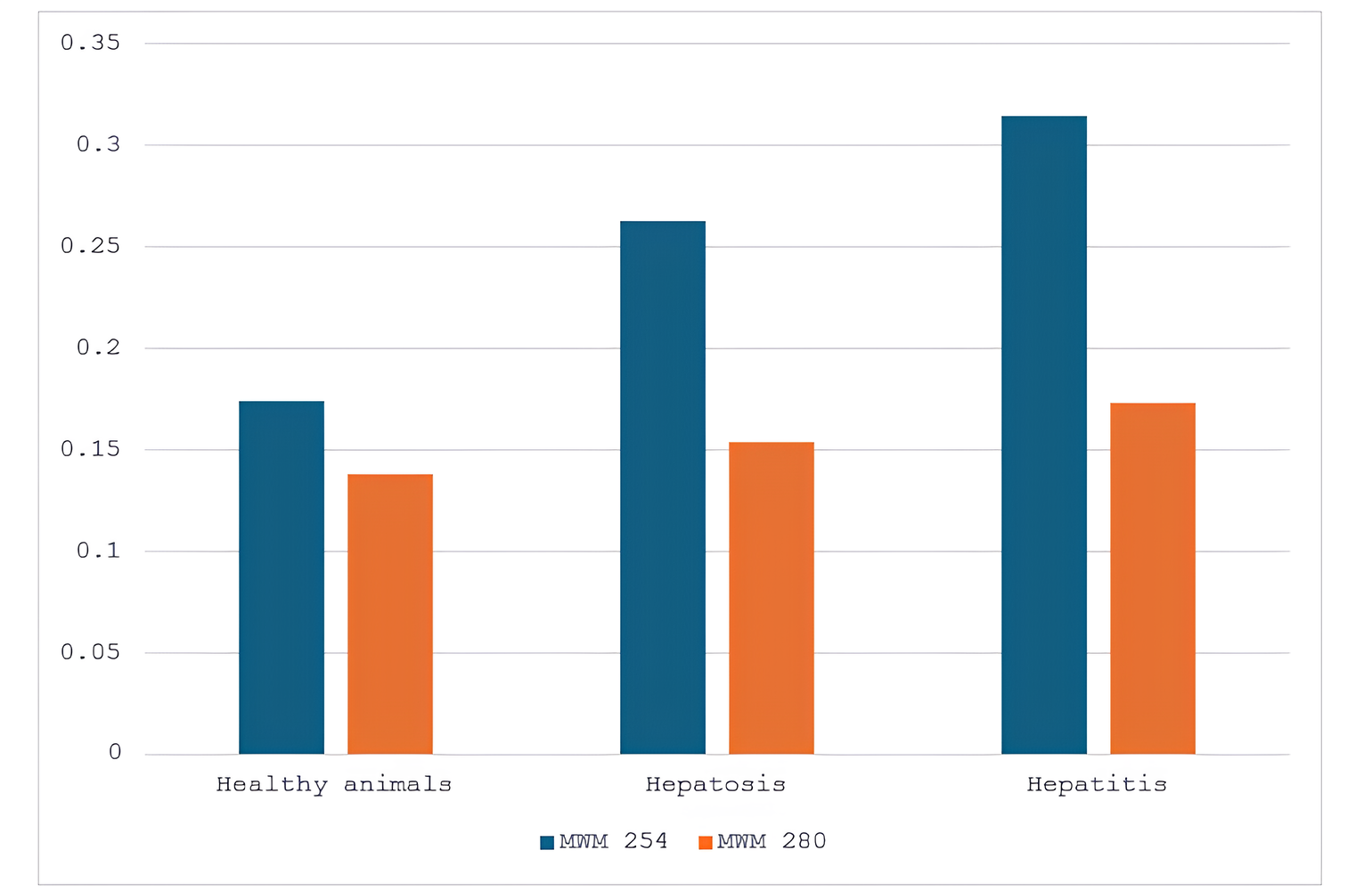
Figure 3 - Fractions of MWM in the blood serum of healthy animals and cows with hepatitis and hepatosis
Foreign authors also propose to evaluate the state of the hepatobiliary system of productive animals by the activity of ornithine transcarbamylase, a mitochondrial enzyme of hepatocytes that catalyzes the reaction between carbamoyl phosphate and ornithine to form citrulline and phosphate , . According to the researchers, this enzyme is a specific indicator of hepatocellular necrosis and fatty liver disease in ruminants. A total of 68 Holstein cattle from different farms in Greece were selected for the experiment. Ornithine transcarbamylase activity in the animals was determined spectrophotometrically. Fatty liver disease was confirmed by histological examination of the organ biopsy. Based on the histological examination results, the experimental animals were ranked into groups according to the following scale:
1 — no morphological changes (there were no animals in this group);
2 — mild fatty hepatosis (4 animals);
3 — moderate fatty hepatosis (10 animals);
4 — moderate to severe fatty hepatosis (different states of hepatocytes were recorded in different lobes of the liver; 13 animals);
5 — severe fatty hepatosis (41 animals) .
The study results are presented below (Table 4).
Table 4 - Ornithine transcarbamylase (OTC), AST and ALT activity in cows with different degrees of fatty hepatosis
Enzymes | Reference values | Animal group | ||||
2 | 3 | 4 | 5 | |||
recovery | death | |||||
OTC, U/l | 2.20-23.60 | 17.03 | 21.31 | 32.40 | 47.01 | 68.06 |
AST, U/l | 26.30-78.90 | 60.50 | 67.85 | 79.73 | 106.52 | 191.97 |
ALT, U/l | 61.00-193.00 | 154.25 | 176.50 | 147.85 | 180.77 | 166.55 |
Note: based on the source [23]
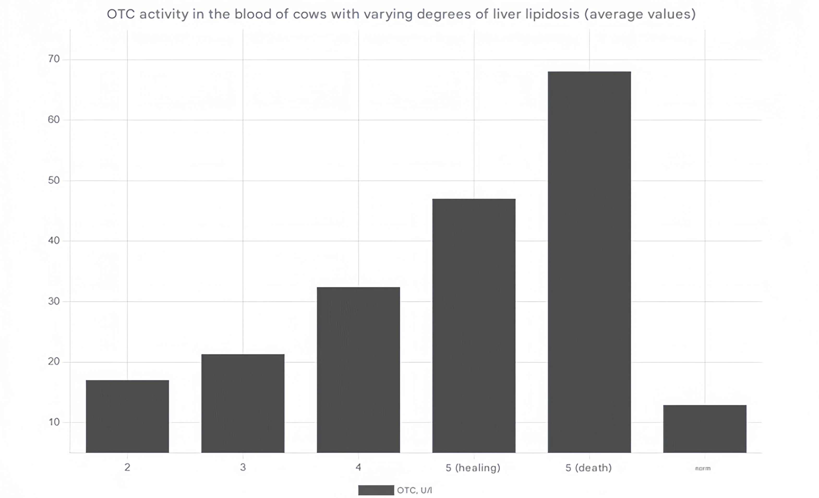
Figure 4 - OTC activity in the blood of productive animals with varying degrees of fatty hepatosis
Some researchers proposed to evaluate the functional state of the liver based on the animal's metabolic profile. It is believed that biological markers of metabolic disorders in high-yielding cows reflect the nature of the processes carried out by the hepatobiliary system. It was found that combined panels of biomarkers have higher diagnostic sensitivity and specificity compared to the analysis of each individual indicator, which allows adapting this method for early diagnostics of fatty hepatosis in productive animals. In experiments , , a search was conducted in dairy cattle for metabolites that met the following criteria:
1) significant differences between the control group and the group with liver pathology;
2) simultaneous conjugate change in direction between the control group and the group with liver pathology.
The feces included two metabolites that satisfied these requirements: behenic acid and L-alpha-aminobutyric acid. 3-nitrotyrosine is one such metabolite that was found in the urine. L-asparagine, palmitoleic acid, L-serine, stearic acid, nonadecanoic acid, petroselinic acid, and heptadecanoic acid were among the pertinent metabolites identified in the blood serum . The animals with hepatic lipidosis had a considerably higher amount of behenic acid and a lower level of the fecal marker L-alpha-aminobutyric acid as compared to the control group. The group of animals with hepatosis had far lower levels of 3-nitrotyrosine in their urine. The content of the remaining five markers (palmitoleic acid, stearic acid, nonadecanoic acid, petroselinic acid, and heptadecanoic acid) exceeded comparable indicators in healthy animals, but only L-asparagine and L-serine showed a significantly lower expression level in the group of cows with liver pathology (Table 5). Changes in the indicated biomarkers are caused by metabolic disorders in dairy cows with fatty hepatosis, in particular, a violation of the metabolism and biosynthesis of arginine, alanine, aspartate, glutamate, proline, unsaturated fatty acids, histidine, cysteine, methionine, pantothenate, coenzyme A, glycine, serine, threonine, taurine, hypotaurine, beta-alanine, and interconversion of pentose and glucuronate in feces .
Table 5 - Potential biomarkers that differ in expression levels in the fatty liver disease group and the control group of animals
Biomarker source | Biomarker | Class | Ratio of biomarker values in animals with liver pathology and healthy cows (path./norm.) |
Blood serum | L-Asparagine | Amino acid | 0.58 |
Palmitoleic acid | Fatty acids | 2.191 | |
L-Serine | Amino acid | 0.579 | |
Stearic acid | Fatty acids | 1.819 | |
Nonadecanoic acid | Fatty acids | 1.678 | |
Petroselinic acid | Organic acids | 2.831 | |
Heptadecanoic acid | Fatty acids | 2.272 | |
Urine | 3-Nitrotyrosine | Amino acid | 0.256 |
Faeces | L-alpha-aminobutyric acid | Amino acid | 0.534 |
Behenic acid | Fatty acids | 1.203 |
Note: based on the source [25]
Thus, the combined analysis of biochemical profile markers for the diagnosis of hepatopathies in productive animals can be successfully integrated and validated in the conditions of modern industrial dairy farming.
A relatively new direction in the diagnosis of functional insufficiency of the hepatobiliary system in productive animals has become the use of dynamic clearance tests, which involve assessing the metabolism of non-toxic exogenous substances for the liver (sorbitol, caffeine, galactose, indocyanine green, lidocaine, and other substances) , . Thus, the studies of Popova O.S. , reflect the key principles of integrating caffeine clearance into the aspect of diagnosing disorders of the hepatobiliary system. In an experiment with cattle, animals from two groups — control (20 animals) and experimental (20 animals with fatty liver disease) — were administered sodium caffeine benzoate at a dosage of 5 mg/kg, and the dynamics of caffeine concentration were observed over 24 hours. The caffeine content in the blood serum was determined by enzyme immunoassay. The obtained results of the study are reflected in the table (Table 6).
Table 6 - Main pharmacokinetic parameters of caffeine clearance in animals of the control and experimental groups: C – control group; E – experimental group
Time after drug administration, h | Caffeine concentration, µg/ml | Total plasma clearance, ml·min⁻¹·kg⁻¹ | ||
C | E | C | E | |
1 | 7.35 | 7.20 | 1.52 | 2.32 |
2 | 6.28 | 6.23 | ||
3 | 5.78 | 5.48 | ||
4 | 4.69 | 4.78 | ||
5 | 3.98 | 3.86 | ||
6 | 3.25 | 3.30 | ||
8 | 2.91 | 2.84 | ||
10 | 2.46 | 2.53 | ||
12 | 1.73 | 1.78 | ||
18 | 0.81 | 1.23 | ||
24 | 0.37 | 0.52 | ||
Note: based on the source [28]
A decrease in caffeine clearance by an average of 7–11% for chronic disorders and by 32-38% for acute disorders directly correlated with a violation of the functional state of the liver, established by standard clinical and biochemical parameters . The principles of using test system models based on measuring the parameters of liver clearance of various inert substances can form the basis of modern approaches to diagnosing hepatobiliary system diseases at early stages.
The pharmacodynamic parameters of indocyanine green are also an effective indicator of general liver function in dairy cattle, since this substance is excreted through the urinary system with less intensity and is therefore more specific for liver damage . Reference values for indocyanine green metabolism in cattle have not yet been established and are currently under investigation. Evaluation of the liver condition using this method is currently possible only through a comparative analysis of the obtained values with the indicators of the control group (healthy animals). It has been experimentally established that the clearance of indocyanine green (ICG) is variable and depends on the periods of feeding and fasting of the animal . Thus, the half-life of ICG has the greatest value after fasting and the smallest 3 hours after feeding (Fig. 5). The authors of the study also found that ICG clearance demonstrated a positive correlation with AST and bilirubin.
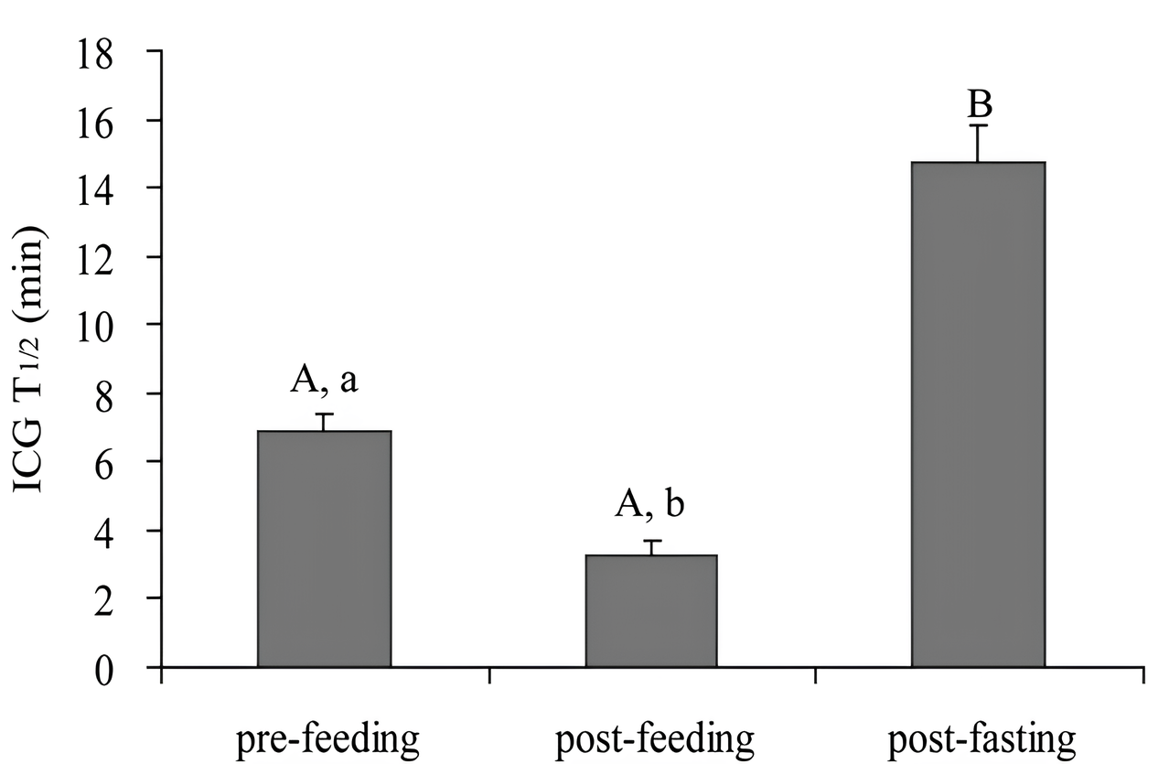
Figure 5 - Half-life (T1/2) values of indocyanine green before feeding, 3 hours after feeding and after fasting in productive animals
Note: based on the source [29]
3. Conclusion
Thus, modern veterinary science has a number of methods for identifying hepatobiliary system pathologies in productive animals , , and , , , . The functional state of the liver can be assessed using various indicators of the animal's biochemical profile, the degree of endogenous intoxication using clearance tests with inert substances, and also through instrumental approaches, such as transient elastography. It should be noted that some approaches require additional studies to establish their effectiveness and reproducibility in clinical settings.