ХАРАКТЕРИСТИКИ МНОГОЯДЕРНЫХ КЛЕТОК В СТРУКТУРЕ МЕЛАНОМЫ ГЛАЗА ЧЕЛОВЕКА
ХАРАКТЕРИСТИКИ МНОГОЯДЕРНЫХ КЛЕТОК В СТРУКТУРЕ МЕЛАНОМЫ ГЛАЗА ЧЕЛОВЕКА

Аннотация
Актуальность данной темы обусловлена тем, что увеальные меланомы относятся к числу наиболее агрессивных опухолей, для которых не существует методов лечения и которые быстро дают метастазы, после чего средняя продолжительность жизни сокращается до 6 месяцев. Высокотехнологичные исследования в основном проводятся на лабораторных животных; экстраполяция данных на человека носит относительный характер. Многоядерные клетки в опухолях (MNTC) описывались многими авторами; однако обзор литературы показал, что MNTC редко встречаются в увеальной меланоме и практически не изучались. Иммуногистохимическое исследование с целью выявления локализации CD68-положительных клеток показало, что MNTC при меланоме сосудистой оболочки глаза не связаны с пулом моноцитов и содержат гетерокарион, который, согласно литературным данным, может влиять на барьерные свойства эпителиального слоя. Из трех известных механизмов слияния клеток наиболее вероятным является вирусная индукция. Дальнейшее изучение MNTC открывает возможности для расширения наших представлений о патогенезе увеальной меланомы.
1. Introduction
Hanratty K., Finegan G., Rochfort K.D., with co-authors (2025), showed that uveal melanoma (UM) is the most common primary intraocular malignant neoplasm in adults worldwide . Shields C.L., Manalac J., Das C., with co-authors (2014), studying primary tumors, Wanten V., Vanhonsebrouck E., Jacob J. (2025), studying secondary metastatic neoplasms, and many other authors, believe that uveal melanoma is a serious intraocular malignant neoplasm that poses a threat to life , . The assessment of clinical characteristics, treatment methods, pathological features, and prognosis of acquired malignant neoplasms of the human choroid, as well as the identification of clinical signs that can help distinguish them from benign ones, is not sufficient to meet the requirements of practical healthcare. According to Kaštelan S., Antunica A.G., Oresković L.B., et al. (2020, 2022), the prognosis for patients with uveal melanoma is unfavorable: the 5-year survival rate is 50–70%, and approximately 50% of patients develop metastases within 15 years of diagnosis, mainly in the liver, with a median survival rate of only 6 months after the appearance of metastases .
The issue of timely enucleation or organ-preserving surgical intervention, as well as the prediction of metastases due to the lack of comprehensive pathogenetically based treatment, remains a matter of debate , . The demonstration of significant clinical differences in each case of uveal melanoma, delayed diagnosis dictate the direction of research to search for additional morphological representations based on human material, since as an extrapolation of molecular genetic data obtained mainly from experimental animals and in vitro tumor cell lines, it has many disadvantages and cannot always be extrapolated to the human body, which indicates the high relevance of research in this area. Information about the pathological anatomy of each case of uveal melanoma in the human eye is invaluable for analysis and application in the development of a personalized treatment strategy.
The aim of the study was to phenotype multinucleated melanoma cells from human choroidal melanoma (CHM) to elucidate their histophysiology and mechanism of formation.
2. Research methods and principles
We studied the material of uveal melanomas from 26 enucleated eyes of patients, as well as 6 cadaveric eyes taken from patients who died from injuries incompatible with life, who were included in the control group. Classical morphological and immunohistochemical methods of study to identify the localization of p53, CD68 and CD163 were performed on all 26 examined melanoma samples in the laboratory of Niigata University (Japan), the neurobiology laboratory of the Far Eastern Federal University; clinical studies and analysis of observational data were carried out at the Primorsky Territory Center for Eye Microsurgery, LLC. Inclusion criteria for the study: age from 29 to 89 years; diagnosed course of the tumor of the uveal duct of varying severity of the clinical course, isolated or in combination with chronic diseases. The classical method of staining with hematoxylin and eosin made it possible to obtain a general morphological picture of uveal melanomas. Immunohistochemistry using DAKO reagents was used to identify the localization of p53, CD68, and CD163-positive cell phenotypes. Section analysis and illustrative material were performed using an Olympus Bx51 microscope and a DP25 digital camera with proprietary software (Japan). This study was conducted in accordance with the fundamental ethical principles of the Declaration of Helsinki and Good Clinical Practice (GCP) and was approved by the Interdisciplinary Ethics Committee of the Far Eastern Federal University, Ministry of Science and Higher Education of the Russian Federation, on June 27, 2024 (Protocol No. 4).
3. Results of own research and their discussion
Analysis of the material revealed that the most common tumors were located in the choroid, less frequently in the region of the ciliary body processes, and most rarely in the iris. Macroscopically, ciliary body tumors predominantly involved the lateral surface of the eye, extending to the posterolateral surface of the choroid. The largest tumor sizes corresponded to the anterior lateral segment of the eye, which led to a delayed diagnosis of melanoma despite gradually increasing clinical manifestations. The tumors were broadly based on the ocular wall, without scleral invasion, with Bruch's membrane protruding toward the vitreous. The topography of five tumors corresponded to a lateral location in the ciliary body, in the area of the blind retina, with extension into the choroid proper. Epithelioid cells were divided into subtypes based on their shape, corresponding to parenchymal epithelial cells, and were classified as tall columnar. In the ciliary body melanoma, epithelioid cells with hypochromic nuclei, eccentric nucleoli, oxyphilic cytoplasm, and clear vesicles located in the apical portion of the cells, along with foamy cytoplasm, were identified. In our study, all tumor cells exhibited an epithelioid/spindle-shaped shape with rare multinucleated cells and nuclear pleomorphism (Figure 1).
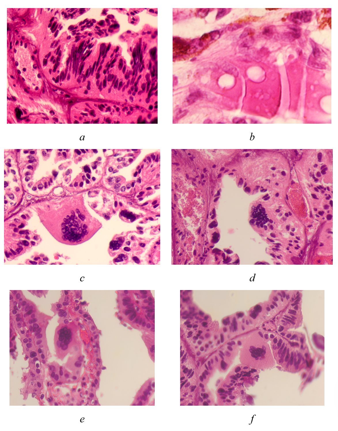
Melanoma of the choroid and ciliary body of the human eye:
a - spindle-shaped cells; b - epithelioid cells with nuclei, devoid of chromatin with vesicular inclusions in the cytoplasm and foamy cytoplasm; c, d, e, f - multinucleated tumor cells
stained with hematoxylin and eosin; magnification A, b x400
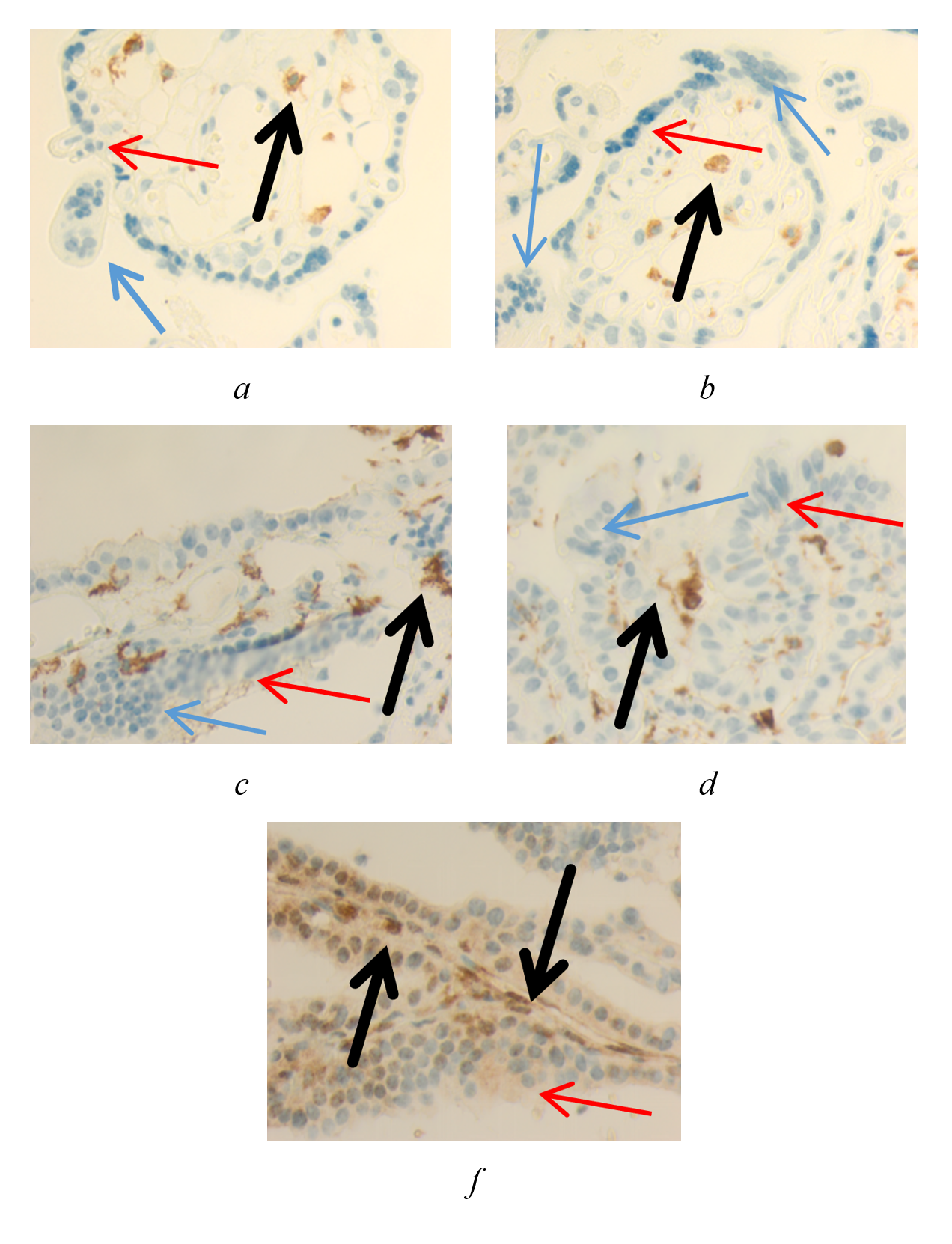
Uveal melanoma of the human eye:
a, b - CD68 positive cell; c, d - CD163 positive cell; f - p53 positive cell
immunohistochemistry to detect the localization of the CD68 marker (a, b); CD163 (c); CD68- and CD163-positive yellow-brown cells are expressed in the stroma of the ciliary processes; p53 (d) is identified in its active (in the nucleus) and inactive (in the cytoplasm) forms; Magnification x200; blue arrows point to multinucleated cells, red arrow points to a developing multinucleated cell, black arrow points to a CD68 (a, b), CD163 (c, d) and p53 (f) positive cell
Indicators of immune competent cells of patients in the observation group in terms of age
differences in data between age groups are statistically significant; p<0.01
Age, years | Number of cells in the field of view (M±m) | ||
CD68 | CD163 | р53 | |
20–39 | 11.17±0.15 | 7.52±0.09 | 3.04±0.07 |
40–59 | 3.44±0.22 | 4.33±0.13 | 11.22±0.08 |
60 and more | 2.31±0.17 | 3.16±0.18 | 15.6±0.12 |
Multinucleated giant cells adjacent to the exposed basement membrane were identified in the examined material. The nuclei were concentrated in the portion of giant macrophages protruding into the vitreous. Multinucleated cells were not identified in all tumors; out of 26, only 14 cases, representing 53.8%, had multinucleated cells.
The absence of CD68 expression as an indicator of phagocytic function in multinucleated cells indicates their unique phenotype and the mechanism of fusion of the original cells.
A virtually complete absence of melanocytes is noted in the area of choroidal malignancy.
The ability of cells to fuse was described by Theodor Schwann (1847), but it was not until 1960 that isolated mouse cells were first intentionally experimentally fused with the same tissue type and their outer membrane fusion induced using Sendai virus (a respiratory virus in mice). This resulted in the formation of a single-nucleated cell with chromosomes from both fusion partners , . Henry Harris of Oxford University (2008) and Nils Ringertz of the Karolinska Institutet in Sweden (2001) showed that proteins from one gene fusion influence gene expression in the nucleus of the other partner, and vice versa , . This changes the cellular phenotype and acquires new functions, including barrier functions. The second type of cell fusion is accompanied by the formation of several nuclei in the cytoplasm, a heterokaryon, which undergo phenotype reversion or transdifferentiation. If nuclear fusion occurs, the fused nucleus initially contains the full chromosomal content of all fusion cytopartners (2Nxn), but eventually the chromosomes are lost and/or reassorted. If nuclear fusion does not occur, the symplast may lose the entire nucleus. This type of heterotypic cell fusion occurs between cells of different types, making it the complete opposite of homotypic cell fusion. Li H., Liu Y., Zeng P. (2025) and Li S., Zou J. (2026) applied an innovative method to formalize multi-stage deep feature fusion with multi-core diversity preservation in omics data and a supervised gate optimization strategy, and developed the scMAG algorithm. This resulted in a framework aimed at improving the accuracy of clustering and visualization of single-cell multi-omics analysis data , .
The result of fusion in our observations is the formation of multinucleated heterokaryons in the absence of nuclear material. This process is analogous to the fusion of the cytoplasm and cytolemma during the formation of bone marrow-derived cell heterokaryons (BMDCs) that combine with cells from other organs. The presence of cells with a nuclear membrane and in the absence of nuclear content, as the most likely outcome of heterokaryon development under conditions of malignancy, is consistent with the results obtained by Ketkar M., Jakati S., Raval V. (2023), characterizing the morphological picture of uveal melanoma with large, oval, vesicular nuclei with the loss of BAP-1 nuclear expression . Zhao T., Denize T., Wang H., et al. (2024) observed multinucleated cells (MNTC) in 15 of 61 cases in tumors at other locations, all of which were negative for CD117. Three tumors underwent chromosomal microarray (CMA) plus gene fusion analysis, as well as FISH and germline testing for alterations in the FLCN and MET genes using PCR, one each. The authors concluded that MNTC are prognostic factors for an unfavorable tumor outcome . Khan Z.M., Cockerell C.J. (1997) discovered multinucleated cells in a skin tumor and identified them as osteoclast-like multinucleated macrophages . Aramin H., Zaleski M., Prieto V.G., Aung P.P. (2019) noted that multinucleated giant cells are rarely found in melanoma , which makes our data particularly valuable due to the fact that their origin and evolution, as well as histophysiological features, remain unknown.
4. Conclusion
The presence of heterokaryons in multinucleated cells during tissue malignancy will expand the range of diagnostic procedures, including those aimed at studying genomic changes, as a platform for creating mathematical prerequisites for determining the developing phenotype of tumor cells. A more comprehensive study of this rare morphological variant of uveal melanoma requires a more in-depth analysis of additional data. Accurate diagnosis will have both prognostic and therapeutic implications. The multinucleated cells we identified do not fit into any of the described classifications of uveal melanomas and currently remain unclassified, requiring further study. Identification of various types of multinucleated giant cells in tumor lesions of the human choroid may aid in the differential diagnosis of these tumors. In all cases, tumors containing multinucleated cells demonstrated a more aggressive course, requiring enucleation. Taken together, the results of this study suggest that the lack of a response to CD68 and CD163 may indicate that the MNTKs do not belong to the monocytic pool. Three known mechanisms of cell fusion—induced by toxins, due to changes in electrical potential, and due to viruses—suggest a more realistic mechanism for heterokaryon formation in this material under viral induction to enhance the barrier properties of the epithelial layer lining the choroidal surface at the anterolateral pole of the eye. This requires further research.