Особенности розеток Флекснера-Винтерштайнера при ретинобластоме
Особенности розеток Флекснера-Винтерштайнера при ретинобластоме

Аннотация
Розетки Флекснера-Винтерштайнера являются структурами, сопровождающими малигнизацию нервной ткани, их роль и функции в канцерогенезе не известны. Наименее изучены Розетки Флекснера-Винтерштайнера при ретинобластоме. С помощью классических и иммуногистохимических методов были изучены розетки Флекснера-Винтерштайнера в структуре глаза 9-летнего мальчика, которому была проведена энуклеация по поводу ретинобластомы. Розетки были обнаружены в сетчатке и стекловидном теле. Представлены характеристики типов клеток в структуре розеток, а также проведен сравнительный анализ типов клеток в зависимости от их расположения и положительной реакции на Ki67. Сделан вывод о важности розеток Флекснера-Винтерштайнера для оценки эффективности лечения и прогнозирования дальнейших стратегий лечения ретинобластомы с целью предотвращения рецидива опухоли и метастазирования.
1. Introduction
A statistical analysis of retinoblastoma (RB) detection data worldwide reveals a steady increase in incidence amid a lack of effective treatment not only for preserving vision but also for preserving organ function. In 2023, 237 papers were published by 351 research institutes from 68 countries and regions, demonstrating the relevance of studying RB .
According to Robertson D.M., Campbell R.J. (1977), of 726 eyes consecutively enucleated at the Mayo Clinic between 1954 and 1974, retinoblastoma was detected in only 41. Despite all efforts aimed at establishing an accurate clinical diagnosis, an erroneous diagnosis of retinoblastoma in a blind eye has much less serious consequences than an erroneous diagnosis of a tumor in a sighted eye . The level of disability and mortality in RB dictates the need to develop a conservative treatment strategy aimed at key pathogenetic targets to replace existing methods with low efficiency, which do not promote full regenerative processes with restoration of visual functions . Ma X., Li X., Sun Q., Luan F., Feng J. (2024), like most authors, believe that retinoblastoma is the most common intraocular malignant tumor in children, arising mainly due to biallelic loss of the RB1 gene in the developing retina . Despite significant progress in understanding the basic pathogenesis of retinoblastoma, a comprehensive study of the complex genetic and epigenetic system underlying the oncogenesis of retinoblastoma remains a serious problem . Traditional treatment methods are limited, and despite the constant identification of genetic loci associated with the pathogenesis of cancer, the development of targeted therapy lags behind the needs of practical healthcare.
The lack of comprehensive data and cellular characteristics of the source of RB for tumor development is one of the pressing issues in modern ophthalmology. Tan C.L., Kimpo M.S., Nga V.D.W., et al. demonstrated the relationship between the RB1 mutation and Flexner-Wintersteiner rosettes in an embryonal tumor and emphasized the need for a large-scale identification of all cases that can help in the clinicopathological correlation of unusual or atypical histological data . Flexner-Wintersteiner rosettes are identified in pathologically altered structures of the eye against the background of RB, the role and functional significance of which in RB is unknown and controversial, their detection is recorded in only 28% of RB, which classifies each case of their detection as unique, which served to determine the direction of our research.
Purpose of the study — to study the localization and prevalence of Flexner-Wintersteiner rosettes in the structures of the human eye in retinoblastoma.
2. Research methods and principles
The study involved ocular material from a 9-year-old boy, enucleated for clinical indications and embedded in paraffin. Material collection was performed in accordance with the provisions of the Declaration of Helsinki, adopted in 2000 and amended in 2013. Three-micron-thick sections prepared from the paraffin block were stained with hematoxylin and eosin to obtain a general morphological picture and immunohistochemistry to identify and localize Ki67-positive cells. Material analysis and image acquisition were performed using an Olympus BX51 microscope and a PDx25 digital camera (Japan).
3. Main results and discussion

Figure 1 - Eye of a 9-year-old boy
Note: Retinoblastoma; Hematoxylin and eosin staining; Microphotograph; Magnification x100
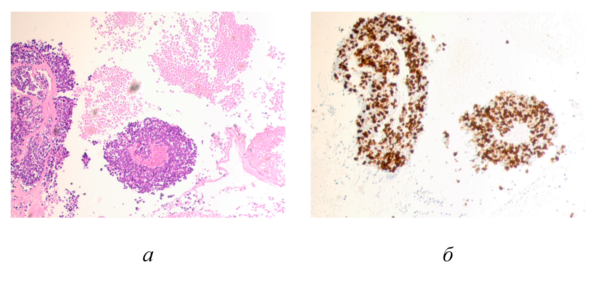
Figure 2 - The eye of a 9-year-old boy with retinoblastoma:
a - hematoxylin and eosin; b - immunohistochemistry to identify the location of Ki67-positive cells
Note: scattered Flexner-Wintersteiner rosettes are observed in the vitreous, most of which are large and multilayered; Photomicrograph; Magnification x100
The Ki-67 index, an index of proliferative activity expressed as a percentage, was 95% in the tumor structure, 42% in the tumor surroundings, and 1% in the marginal zone. Low index values in the tumor surroundings indicate a less aggressive malignancy and tumor growth process, but a high degree of aggressiveness in the structure of the Flexner-Wintersteiner rosettes. The classic Flexner-Wintersteiner rosette is a rounded cluster of cells grouped around a central lumen containing small cytoplasmic processes of the surrounding cells. The proliferative activity index demonstrated tolerance of the ocular structures to the treatment and the absence of an effective tumor response to conservative therapy. The data obtained predict rapid tumor growth and spread to all ocular structures, making eye extirpation the most appropriate strategy for stopping the malignancy. The evidence that Ki67-positive cells occupy the majority of the malignant tissue of Flexner-Wintersteiner rosettes, while the proliferative activity indices are lower at the periphery, does not contradict the data obtained by other authors that the malignancy process occurs against the background of possible partial differentiation of cells in the tumor zone towards photoreceptor cells. Electron microscopy performed by Karim M.M., Yamamoto M., Itoh H. (1996) showed that tumor cells forming the Flexner-Wintersteiner rosette have ultrastructural features of primitive photoreceptor cells . Transmission electron microscopy revealed the presence of large mitochondria on the luminal side of the cells forming the rosettes, which are presumably part of the inner segments of photoreceptor cells. The results of this study indicate a predominantly neuronal nature of neoplastic cells with differentiation characteristic of photoreceptors. Tajima Y., Munakata S., Ishida Y., et al. (1994) observed an increase in the number of long mitochondria and microtubules in the cell cytoplasm, indicating differentiation towards photoreceptors. The authors also noted that hereditary retinoblastoma demonstrated a higher degree of cell differentiation than the non-hereditary type. Differentiated photoreceptor retinoblastoma grew more slowly compared to undifferentiated retinoblastoma, so with the first variant, one can count on a cure with surgery . These observations allow us to draw additional conclusions about the biological nature of retinoblastomas. In addition, the cells in the rosette structure are stained in the same way as rods and cones, which allowed the authors to suggest that Flexner-Wintersteiner rosettes represent a special form of cellular differentiation of the retina. Flexner-Wintersteiner rosettes are characteristic not only of retinoblastomas, but also of pineoblastomas and medulloepitheliomas. Our data indicating that cells in the Flexner-Wintersteiner rosette structure, located in an area with a high concentration of Ki67-positive cells and undergoing irreversible mitosis, indicate a lack of differentiation and specialization of cells entering mitosis. The lumen filled with cells corresponding to blood cells indicates that Flexner-Wintersteiner rosettes in the vitreous body are formed due to angiogenesis and blood infiltration. When culturing retinoblastoma, Bogenmann E. and Mark C. (1983) consistently identified the spontaneous formation of well-differentiated Flexner-Wintersteiner rosettes, provided these structures were present in the patient's primary eye tumor .
The mechanism for inducing high expression of Ki67-positive cells is associated with mutations in the RB1 gene, which lead to complete suppression of retinoblastoma protein (pRb) secretion. The tumor suppressor protein pRb is a key inhibitor of cell proliferation, regulating cell cycle transition to the S phase by blocking the G1 checkpoint. Blocking the G1/S transition occurs primarily through interaction with transcription factor 1 of the E2F family. Mutations in the pRb protein result in activation of the E2F1 transcription factor, resulting in uncontrolled cell proliferation and the formation of tumor cell clones.
4. Conclusion
However, a wider range of genetic and epigenetic alterations that may influence RB1, with further different clinical outcomes, should be considered. According to Roohollahi K., de Jong Y., van Mil S.E., Fabius A.W.M., Moll A.C., Dorsman J.C. (2022), there are other mutations, such as MYCN amplification, that contribute to the formation of particularly aggressive tumors that are completely independent of RB1 . Xu M., Shi H., Shen Y., et al. (2025) studied the materials on cavernous retinoblastoma associated with improved metastasis-free survival (P = 0.007) and overall survival (P = 0.03), as well as with an increase in the proportion of well-differentiated status (P < 0.001) and a decrease in the frequency of metastasis to the vitreous body (P = 0.02), established a new genetic-phenotypic relationship, showing that it is the decrease in MYCN expression that induces the formation of translucent cavities in RB with the phenotype of a less aggressive, well-differentiated RB subtype with a more favorable prognosis . Conclusion. The heterogeneity and cellular compositional features of Flexner-Wintersteiner rosettes and their environment, characteristic of retinoblastoma at the molecular, cellular, and tissue levels in its current models, will allow the creation of promising platforms that could contribute to the development of effective patient-specific RB treatment strategies, as well as the choice of further treatment method for patients after enucleation to prevent the progression of tumor spread along the optic nerve and further metastasis. This opinion is also shared by Ryl T., Afanasyeva E., Hartmann T., et al. (2024); Liao Q., Yang J., Shi H., (2025), who demonstrated that inhibition of MYCN activity can restore more differentiated and less aggressive tumor biology , . Therefore, Flexner-Wintersteiner rosettes can serve as potential therapeutic targets for the treatment of retinoblastoma and research in the field of targeted therapy. Although rare, Flexner-Wintersteiner rosettes can serve as a reliable diagnostic feature of RB. Cytomorphologically, tumor cells within Flexner-Wintersteiner rosettes are divided into two types, corresponding to their histological degree of differentiation. These features are cytodiagnostic markers of retinoblastoma and can assist in establishing a cytological diagnosis, assessing the effectiveness of treatment, and predicting further treatment strategies.