Клеточные и молекулярные механизмы ангиогенеза в опухолях нервной и сенсорной систем
Клеточные и молекулярные механизмы ангиогенеза в опухолях нервной и сенсорной систем
Аннотация
Актуальность. Несмотря на известные ангиогенные рецепторы и факторы, активирующиеся в опухолях, стимулирующие сигнальные пути ангиогенеза подавлением генов-супрессоров опухолей в различных органах, этот вопрос остается наименее изученным в малигнизирующихся структурах глаза человека
, . Экспрессия VEGF и CD34 может служить важным прогностическим биомаркером в исходе опухолей мозга и сенсорных систем, а также при разработке стратегии ангиогенного таргетного лечения , . Высокая смертность пациентов с опухолями нервной и сенсорных систем диктуют направление исследований на изучение ключевых мишеней для повышения эффективности таргетной антиангиогенной терапии, снижающей рост опухоли , , что послужило для выбора нами направления исследований.Цель исследования: установить особенности локализации и уровень экспрессии VEGFA и CD34 – позитивных клеток в опухолях мозга и сосудистой оболочки глаза человека для разработки таргетного консервативного лечения с контролируемым прогнозом исхода.
Материалы и методы: с помощью иммуногистохимического метода на выявление уровня экспрессии VEGFА и CD34 изучены 41 биоптат опухолей мозга и 15 биоптатов опухолей глаза человека.
Результаты исследования: установлено, что на фоне ангиогенеза в опухолях мозга и цилиарного тела глаза человека часть сосудов подвержена кальцификации и облитерации просвета с образованием псаммозных телец. Идентификация псаммом в строме цилиарного тела и в стекловидном теле свидетельствует о давности развития процесса малигнизации.
VEGFА позитивность экспрессируется в высокой степени в беспигментном эпителии отростков цилиарного тела и опухоли мозга. CD34 маркером оценён ангиогенез на фоне развивающихся злокачественных опухолей.
Заключение: отсутствие пигмента в эпителии отростков цилиарного тела и локализация высокой экспрессии ангиогенных факторов в стекловидном теле свидетельствуют о соответствии процесса малигнизации в сосудистой оболочке глаза эмбриональному гистогенезу. Таргетная антиангиогенная терапия с помощью ингибиторов ангиогенеза может быть направлена на строму цилиарных отростков и в стекловидное тело в соответствии с локализацией опухоли. Уровень экспрессии VEGFА и CD34 позитивных клеток коррелирует с деструктивными процессами в опухолях и окружающих опухоли тканях.
1. Introduction
In tumor histogenesis, the level of aggressiveness and the tendency to metastasize are affected by angiogenesis, which develops under the influence of increasing hypoxia
, , which has been known for more than a century since Rudolf Virchow obtained evidence of the proliferation of blood vessels in a developing tumor and its dependence on angiogenesis, which is induced by the tumor cells themselves , . Angiogenesis is a complex process, accompanied, according to Ahir B.K., Engelhard H.H., Lakka S.S. (2020), by proliferation, migration and differentiation of endothelial cells under the influence of specific signalling molecules with the control of inducers and inhibitors , which has been studied to a greater extent in experimental animals and requires in-depth study on human material. This fact served as a basis for our choice of research direction.2. Material and methods
A total of 41 biopsies of primary brain tumors and postoperative material from the removal of 15 melanomas of the ciliary body of the eye of patients of different ages and sexes were studied. Sections of 5 μm thickness were made from paraffin blocks, followed by staining with hematoxylin and eosin and examined using the immunohistochemical method to identify the localization and level of expression of VEGFА and CD34.
The relationship between the microvessel density (MVD), the expression of the vascular endothelial growth factor (VEGF) and histopathology in primary tumors of the brain and ciliary body was compared. The removed tumors were examined for MVD using immunostaining with CD34 antibodies, as well as VEGF expression, assessed using immunostaining with VEGF antibodies.
Microscopic analysis of the preparations was performed on an Olympus Bx52 microscope. Histology slide images were acquired using CellSens proprietary imaging software (Olympus Life Science, Tokyo, Japan), and digital slides were prepared using Aperio Scan Scope (Leica Biosystems Inc., Buffalo Grove, IL).
3. Research results and their discussion
A study of sections of human eye tumor biopsies showed the presence of psammosis bodies of various shapes and sizes in the stromal tissue of the ciliary body of the human eye, identical to calcifications in brain tumors (Fig. 1).
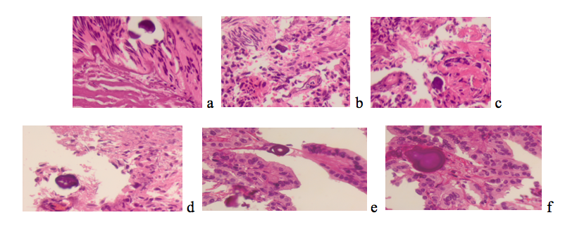
Figure 1 - Processes of the ciliary body of the human eye. Psammosis bodies. Hematoxylin and eosin staining
Note: microphoto. Magnification X400
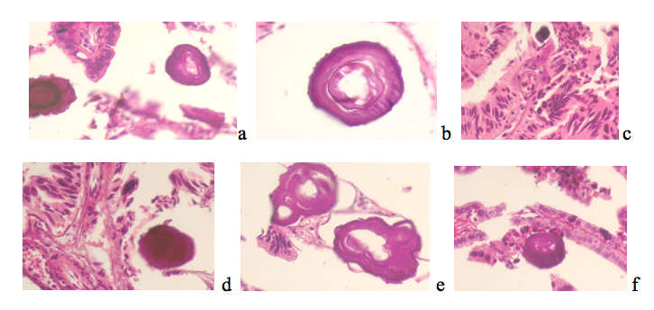
Figure 2 - Psammosis bodies identified in the ciliary body, vitreous body of patients with ocular melanoma. Psammosis bodies. Hematoxylin and eosin staining
Note: microphoto. Magnification X400
We have established that some bodies had unstained lumens in the central part, a round or oval shape, a layered structure, compacted areas in the wall of psammous bodies and sizes from 5 to 15 μm. Analysis of the obtained material allowed us to note both complete obliteration of blood vessels and the preservation of a small lumen inside the vessel, as a result of incomplete calcification. The process of vascular calcification and the absence of vascular endothelium on the inner surface were previously called vasculogenic mimicry – the ability of tumor cells to form vascular-like channels limited by the basal membrane without the participation of endothelial cells and fibroblasts. Vasculogenic mimicry (VM) was first discovered by R. Folberg et al. (1992) in the pathohistological material of patients with choroidal melanoma and was subsequently confirmed in vitro during the cultivation of tumor cells on extracellular matrices
. The development of vasculogenic mimicry is a process of vascular calcification with the formation of psammous bodies (PB), a complex biological process involving several signalling pathways. The fact that VM-PB is found in various types of aggressive tumors (in melanoma of the ciliary body and brain tumors) indicates that this process reflects the characteristics of tumor aggressiveness. More and more experimental data are accumulating indicating a high statistical correlation between the presence of PB and the ability of the tumor to metastasize . The presence of unchanged blood vessels with a typical structure for arterioles located near PB suggests that calcification primarily affects the vessels of the ciliary body outflow system (Fig. 3).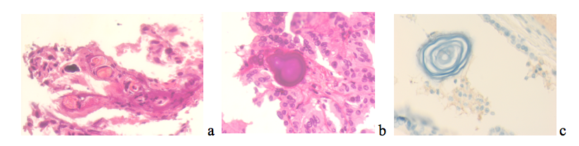
Figure 3 - Psammosis bodies in the ciliary body processes of the human eye
Note: a, b – hematoxylin and eosin staining; c – immunohistochemistry to detect VEGFA positive expression.
Microphoto. Magnification X400
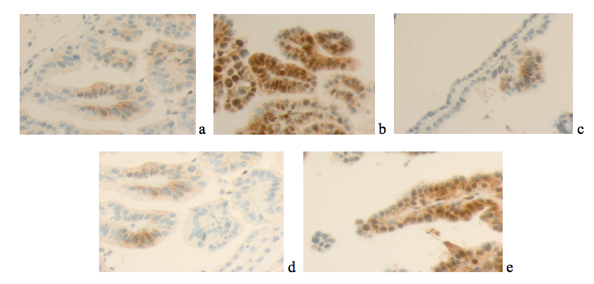
Figure 4 - Processes of the ciliary body of the human eye. Immunohistochemistry. Localization of VEGFА positive expression
Note: microphoto. Magnification X400
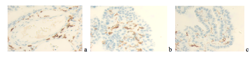
Figure 5 - Ciliary body processes of the human eye. Immunohistochemistry. Localization of CD34 positive expression
Note: microphoto. Magnification X400
4. Conclusion
The absence of pigment in the epithelium of the ciliary body processes and the localization of high expression of angiogenic factors in the vitreous body around the ciliary body processes, in the ciliary epithelium, and not only in the vascular endothelium, indicate that the malignancy process in the choroid of the human eye corresponds to embryonic histogenesis using signalling pathways of early human ontogenesis. Targeted antiangiogenic therapy using angiogenesis inhibitors can be directed not only to the stroma of the ciliary processes, but also directly to the vitreous body of the human eye in the anterior right and left sectors of the eye and in accordance with the localization of the tumor. The level of expression of VEGFА and CD34 positive cells correlates with destructive processes in tumors and surrounding tissues. Analysis of the identified features of localization of CD34 and VEGFA positive expression, molecular mediators that regulate angiogenesis in malignant tissues of the brain and ciliary body of the human eye, contributes to the disclosure of the mechanisms of tumor angiogenesis and dictates the direction of the vector of clinical studies for the development of a strategy for the conservative treatment of glioblastomas and ciliary body tumors using angiogenic pathways and corresponding signaling molecules.