ИССЛЕДОВАНИЕ МИКРОЦИРКУЛЯЦИИ КРОВИ В КОЖЕ РАЗЛИЧНЫХ ОБЛАСТЕЙ ТЕЛА МЕТОДОМ ЛДФ
Козлов В.И.1, Гурова О.А.2
1Доктор медицинских наук, профессор,
2Кандидат биологических наук, доцент,
Российский университет дружбы народов, Москва
ИССЛЕДОВАНИЕ МИКРОЦИРКУЛЯЦИИ КРОВИ В КОЖЕ РАЗЛИЧНЫХ ОБЛАСТЕЙ ТЕЛА МЕТОДОМ ЛДФ
Аннотация
С помощью лазерной допплеровской флоуметрии (ЛДФ) изучали параметры микроциркуляции крови в коже различных топографо-анатомических областей тела у здоровых лиц в возрасте 18-24 лет. Особенности строения кожи и ее микроциркуляторного русла в этих участках тела исследованы гистологическими методами и с помощью капилляроскопии. Определены нормативные показатели состояния микроциркуляции крови в коже головы, туловища и основных сегментов верхней и нижней конечностей. Величина параметров ЛДФ в коже разных областей тела зависит от плотности функционирующих капилляров, толщины эпидермиса и глубины залегания микрососудов. Ключевые слова: ЛДФ, микроциркуляция крови, области тела.Kozlov V.I.1, Gurova O.A.2
1MD, Full Professor,
2PhD in biological sciences, associate Professor,
Peoples’ Friendship University of Russia, Moscow
THE STUDY OF BLOOD MICROCIRCULATION IN THE SKIN OF DIFFERENT AREAS OF THE BODY BY LDF
Abstract
Using laser Doppler flowmetry (LDF) studied parameters of the skin microcirculation of different topographo-anatomic regions of the body at healthy persons age of 18-24 years. Features of the structure of the skin and microvessels in these areas of the body are investigated by histological methods and capillaroscopy. Normative parameters of a condition of microcirculation in a skin of head, body and main segments of hand and leg are determined. The magnitude of the parameters of LDF affect the density of functioning capillaries, the thickness of epidermis and the depth of the microvasculature in the skin. Keywords: LDF, skin microcirculation, regions of the body. Assessment of the state of microcirculation in different body areas is of great importance in clinical practice and research. Method of laser Doppler flowmetry (LDF) is a modern, effective and non-invasive method of microcirculation diagnostics [3-5]. However, until recently, regulatory figures of LDF-grams of the skin of different topographic areas of the body have not been clearly defined. Moreover their dependence on the specific structure of the microcirculation in the skin of these areas have not been studied [1, 2]. The purpose of this study was to determine the values of LDF of the skin of different body areas in healthy young people, and demonstrate the peculiarities of microcirculation of skin in these areas. Material and methods of research Morpho-functional peculiarities of blood microcirculation were studied in the skin of 15 anatomical areas of the body in 80 healthy males aged 18-24 years. Morphological and functional characteristics were recorded in the skin of the breast (5th intercostal space, right anterior axillary line), abdomen (lateral edge of rectus abdominis muscle), forehead, ear lobule, arm (the medial surface of the lower third), forearm (ventral surface of the lower third), hand (1st dorsal interosseous space), ring finger (palmar and the dorsal surface of the distal phalanx), hip (the medial surface of the lower third), leg (posterior surface of the lower third), medial and lateral malleoli, foot (1st dorsal interosseous space), great toe (posterior surface of the distal phalanx). The microcirculation status was evaluated using laser analyzer of blood flow "LAKK-01" ("Lazma", Russia) in subjects sitting in accordance with the methodological recommendations [4]. Biomicroscopic study of skin capillaries was performed by microscope MLK-3MT (LOMO) with the use of the capillaroscopy method . Histological methods (standard staining of the skin with hematoxylin and subsequent eosin) revealed the depth of blood vessels in the same skin area of cadaver. All data was processed by the methods of variation statistics. Results of the study Blood microcirculation figures in the skin of different body areas, obtained by LDF method, are presented in table 1.Table 1 - Parameters of blood microcirculation in the skin of the different areas of the body
| The area of the body | PM | SD | IF |
| Forehead | 18,7±0,5 | 1,8±0,1 | 1,18±0,02 |
| Ear lobule | 27,9±1,2 | 3,3±0,3 | 1,56±0,04 |
| Breast | 14,6±1,0 | 2,0±0,2 | 1,53±0,05 |
| Abdomen | 12,4±0,6 | 1,2±0,1 | 1,51±0,04 |
| Arm | 10,1±1,0 | 1,2±0,1 | 1,65±0,05 |
| Forearm | 6,7±0,3 | 0,8±0,1 | 1,63±0,05 |
| Hand | 7,1±0,3 | 1,2±0,1 | 1,94±0,07 |
| Finger (palmar surface) | 25,4±0,9 | 3,1±0,2 | 2,09±0,05 |
| Finger (dorsal surface) | 17,1±1,0 | 2,9±0,2 | 1,89±0,06 |
| Hip | 6,5±0,3 | 0,7±0,03 | 1,67±0,06 |
| Leg | 6,1±0,3 | 0,5±0,04 | 1,54±0,06 |
| Medial malleolus | 6,1±0,2 | 0,6±0,03 | 1,57±0,06 |
| Lateral malleolus | 6,7±0,2 | 0,6±0,04 | 1,57±0,05 |
| Foot | 7,2±0,4 | 0,6±0,1 | 1,53±0,05 |
| Great toe (dorsal surface) | 6,9±0,4 | 0,8±0,1 | 1,59±0,06 |
Table 2 - Spectral characteristics of oscillations of skin blood flow in the skin of different anatomical areas of the body (contribution of frequency components to the total spectrum power in %)
| The area of the body | Frequency components | |||
| VLF | LF | HF | CF | |
| Forehead | 50,1±2,21 | 37,9±2,4 | 9,2±0,67 | 2,8±0,3 |
| Ear lobule | 53,0±2,33 | 35,4±2,56 | 9,2±0,99 | 2,4±0,42 |
| Breast | 44,5±1,16 | 42,5±0,98 | 10,9±0,69 | 2,13±0,28 |
| Abdomen | 52,2±0,48 | 39,8±0,36 | 6,4±0,29 | 1,6±0,11 |
| Arm | 49,9±1,19 | 40,4±0,98 | 8,6±0,63 | 1,1±0,25 |
| Forearm | 50,6±1,08 | 39,3±1,06 | 9,1±0,65 | 1,0±0,09 |
| Hand | 53,2±2,01 | 39,6±1,77 | 6,2±0,35 | 1,0±0,14 |
| Finger (palmar surface) | 54,1±0,78 | 38,4±0,35 | 6,6±0,42 | 0,9±0,07 |
| Finger (dorsal surface) | 56,4±0,89 | 36,8±0,61 | 6,1±0,29 | 0,7±0,06 |
| Hip | 50,9±0,73 | 40,1±0,52 | 7,6±0,58 | 1,4±0,15 |
| Leg | 51,4±0,92 | 37,9±0,84 | 9,2±0,77 | 1,5±0,15 |
| Medial malleolus | 49,8±1,01 | 40,9±0,88 | 8,1±1,07 | 1,2±0,12 |
| Lateral malleolus | 51,8±2,06 | 36,5±2,15 | 9,8±0,85 | 1,9±0,21 |
| Foot | 50,3±0,96 | 38,8±0,48 | 9,1±0,86 | 1,8±0,45 |
| Great toe (dorsal surface) | 54,8±1,57 | 35,2±1,27 | 8,7±0,79 | 1,3±0,18 |
Table 3 - Features of the structure of the skin in different anatomical areas of the body
| The area of the body | Density functional capillaries1 mm2 | Thickness epidermis, mcm | Depth | |
| capillaries papillary layer, mcm | vessels of subpapillar plexus, mcm | |||
| Forehead | 28±0,4 | 52±0,4 | 55±0,35 | 87±0,35 |
| Ear lobule | 54±0,6 | - | - | - |
| Breast | 16±0,5 | 53±0,45 | 56±0,35 | 89±0,45 |
| Abdomen | 14±1 | 38±0,6 | 49±0,4 | 106±0,95 |
| Arm | 12±0,4 | 74±0,71 | 78±1,21 | 123±0,75 |
| Forearm | 16±0,4 | 72±1,31 | 83±1,11 | 121±0,85 |
| Hand (dorsal surface) | 48±0,5 | 88±0,85 | 89±0,85 | 133±0,65 |
| Hand (palmar surface) | - | 516±2,32 | 530±1,26 | 619±0,55 |
| Ring finger (dorsal surface) | 57±0,7 | 131±2,52 | 94±0,65 | 167±1,11 |
| Hip | 14±0,8 | 62±0,95 | 66±0,7 | 85±0,45 |
| Leg | 16±0,6 | 129±1,11 | 136±0,8 | 147±0,55 |
| Medial maleolus | 22±0,5 | 146±0,9 | 149±0,7 | 174±0,6 |
| Foot (dorsal surface) | 36±0,6 | 138±0,95 | 145±0,85 | 167±0,8 |
| Foot (plantar surface) | - | 631±2,47 | 652±1,71 | 727±1,26 |
| Great toe (dorsal surface) | 37±0,5 | 144±0,7 | 150±1,31 | 188±0,96 |
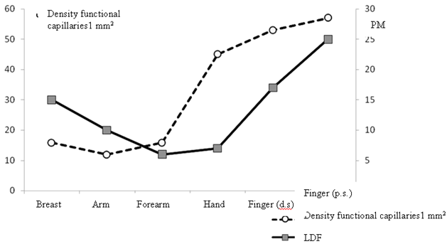
Fig. 1 - The ratio of the density of capillaries and microcirculation in the skin of the breast and different parts of the upper limb
The reduction of PM was observed in the skin of the proximal parts of the extremities (shoulder and thigh) in comparison with the figures obtained in the skin of the trunk together with the trend of gradual increase of the index closer to the distal parts of the limbs (Fig. 1). In the skin of the upper extremity this pattern is expressed more vividly: the value of PM in the skin of the finger significantly (p ≤ 0,01) surpasses those in the skin of other parts of the extremity. Thus, the value of the baseline LDF increases correspondingly with the relocation of the examined area from the body to the distal parts of the upper extremity. That correlates with the increase of capillaries density in the skin of these areas. Low values of LDF-grams in the skin of the different parts of the lower extremities are caused by the depth of location of the microvessels and the influence of the position of the lower limbs.References
- Kozlov V.I. System of microcirculation: clinical and morphological aspects of research // Regional circulation and microcirculation. - 2006. – T.5, № 2. - P.84-101.
- Kozlov V.I. Development of the system of microcirculation.-M: RUDN, 2012.-314 p.
- Intaglietta M. Capillary Flow motion // J. Microcirculation. - 2002-Vol.14 (suppl.l.) - P. 3 - 15.
- Krupatkin A.I., Sidorov V.V. Laser Doppler flowmetry blood microcirculation.- M: Medicine, 2005. - 254 p.
- Fargell B. Problems using laser Doppler on the skin in clinical practice. // Laser Doppler. - London, Los Angeles, Nicosia. - Med-Orion Publish. Co., 1994. - P. 49 - 54.